Home > Product > Antibody > Mouse Anti-FGFR1 antibody
bFGF R; BFGFR; C FGR; CD 331; CD331; CD331 antigen; CEK; FGFBR; FGFR 1; FGF Receptor 1; Fibroblast growth factor receptor 1; FLG; FLG protein; FLJ14326; FLT 2; FLT2; Fms like tyrosine kinase 2; Fms related tyrosine kinase 2; Fms related tyrosine kinase 2
Cat:
SLM51288M
Species Reactivity:
Human,
Immunogen:
KLH conjugated synthetic peptide derived from human FGFR1:718-822/822
Format:
Liquid
Storage instructions:
Shipped at 4℃. Store at -20 °C for one year. Avoid repeated freeze/thaw cycles.
Concentration:
1mg/ml
Clonality:
Monoclonal
Isotype:
IgG1,k
Applications:
WB=1:500-2000IHC-P=1:20-100IHC-F=1:20-100IF=1:20-100(Paraffin sections need to do antigen repair)not yet tested in other applications.optimal dilutions/concentrations should be determined by the end user.
Host:
Mouse
Product Overview:
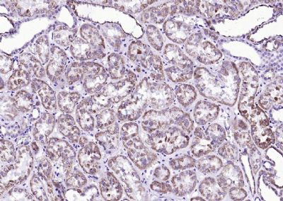
Paraformaldehyde-fixed, paraffin embedded (Human kidney); Antigen retrieval by boiling in sodium citrate buffer (pH6.0) for 15min; Block endogenous peroxidase by 3% hydrogen peroxide for 20 minutes; Blocking buffer (normal goat serum) at 37°C for 30min; Antibody incubation with (FGFR1) Monoclonal Antibody, Unconjugated (SLM51288M) at 1:200 overnight at 4°C, followed by operating according to SP Kit(Mouse)(sp-0024) instructionsand DAB staining.
More
Unit:
Price: $
Product PDFs
Datasheet:


Fibroblast growth factors (FGFs) produce mitogenic and angiogenic effects in target cells by signaling through the cellular surface tyrosine kinase receptors. There are four members of the FGF receptor family: FGFR-1 (flg), FGFR-2 (bek, KGFR), FGFR-3 and FGFR-4. Each receptor contains an extracellular ligand binding domain, a transmembrane region and a cytoplasmic kinase domain (1). Following ligand binding and dimerization, the receptors are phosphorylated at specific tyrosine residues (2). Seven tyrosine residues in the cytoplasmic tail of FGFR-1 can be phosphorylated: Tyr463, Tyr583, Tyr585, Tyr653, Tyr654, Tyr730 and Tyr766. Tyrosine 653 and 654 are important for catalytic activity of the activated FGFR and are essential for signaling (3). The other phosphorylated tyrosine residues may provide docking sites for downstream signaling components such as Crk and PLCgamma.

Function:
Receptor for basic fibroblast growth factor. Receptor for FGF23 in the presence of KL. A shorter form of the receptor could be a receptor for FGF1 (aFGF).

Subunit:
Monomer. Homodimer after ligand binding. Interacts predominantly with FGF1 and FGF2, but can also interact with FGF3, FGF4, FGF5, FGF6, FGF8, FGF10, FGF19, FGF21, FGF22 and FGF23 (in vitro). Ligand specificity is determined by tissue-specific expression of isoforms, and differences in the third Ig-like domain are crucial for ligand specificity. Affinity for fibroblast growth factors (FGFs) is increased by heparan sulfate glycosaminoglycans that function as coreceptors. Likewise, KLB increases the affinity for FGF19, FGF21 and FGF23. Interacts (phosphorylated on Tyr-766) with PLCG1 (via SH2 domains). Interacts with FRS2A. Interacts (via SLCterminus) with NEDD4 (via WW3 domain). Interacts with KL. Interacts with SHB (via SH2 domain) and GRB10. Interacts with KAL1; this interaction does not interfere with FGF2-binding to FGFR1, but prevents binding of heparin-bound FGF2. Interacts with SOX2 and SOX3

Subcellular Location:
Cell membrane; Single-pass type I membrane protein. Nucleus. Cytoplasm, cytosol. Cytoplasmic vesicle. Note=After ligand binding, both receptor and ligand are rapidly internalized. Can translocate to the nucleus after internalization, or by translocation from the endoplasmic reticulum or Golgi apparatus to the cytosol, and from there to the nucleus.

Tissue Specificity:
Detected in astrocytoma, neuroblastoma and adrenal cortex cell lines. Some isoforms are detected in foreskin fibroblast cell lines, however isoform 17, isoform 18 and isoform 19 are not detected in these cells.

Post-translational modifications:
Binding of FGF1 and heparin promotes autophosphorylation on tyrosine residues and activation of the receptor.

DISEASE:
Defects in FGFR1 are a cause of Pfeiffer syndrome (PS) ; also known as acrocephalosyndactyly type V (ACS5). PS is characterized by craniosynostosis (premature fusion of the skull sutures) with deviation and enlargement of the thumbs and great toes, brachymesophalangy, with phalangeal ankylosis and a varying degree of soft tissue syndactyly.
Defects in FGFR1 are a cause of idiopathic hypogonadotropic hypogonadism (IHH) [MIM:146110]. IHH is defined as a deficiency of the pituitary secretion of follicle-stimulating hormone and luteinizing hormone, which results in the impairment of pubertal maturation and of reproductive function.

Similarity:
Belongs to the protein kinase superfamily. Tyr protein kinase family. Fibroblast growth factor receptor subfamily.
Contains 3 Ig-like C2-type (immunoglobulin-like) domains.
Contains 1 protein kinase domain.

SWISS:
P11362

Gene ID:
2260

Database links:

Entrez Gene: 2260 Human

Entrez Gene: 14182 Mouse

Entrez Gene: 79114 Rat

Omim: 136350 Human

SwissProt: P11362 Human

SwissProt: P16092 Mouse

SwissProt: Q04589 Rat

Unigene: 264887 Human

Unigene: 265716 Mouse

Unigene: 207203 Rat

Unigene: 9797 Rat


Picture

Paraformaldehyde-fixed, paraffin embedded (Human kidney); Antigen retrieval by boiling in sodium citrate buffer (pH6.0) for 15min; Block endogenous peroxidase by 3% hydrogen peroxide for 20 minutes; Blocking buffer (normal goat serum) at 37°C for 30min; Antibody incubation with (FGFR1) Monoclonal Antibody, Unconjugated (SLM51288M) at 1:200 overnight at 4°C, followed by operating according to SP Kit(Mouse)(sp-0024) instructionsand DAB staining.
Product Feedback Wall
Message :
Your Email :
Copyright © 2007-2018 Sunlong Medical All Rights Reserved.