This gene is a member of the TIMP gene family. The proteins encoded by this gene family are natural inhibitors of the matrix metalloproteinases, a group of peptidases involved in degradation of the extracellular matrix. In addition to an inhibitory role against metalloproteinases, the encoded protein has a unique role among TIMP family members in its ability to directly suppress the proliferation of endothelial cells. As a result, the encoded protein may be critical to the maintenance of tissue homeostasis by suppressing the proliferation of quiescent tissues in response to angiogenic factors, and by inhibiting protease activity in tissues undergoing remodelling of the extracellular matrix. [provided by RefSeq, Jul 2008].
Function:
Complexes with metalloproteinases (such as collagenases) and irreversibly inactivates them by binding to their catalytic zinc cofactor. Known to act on MMP-1, MMP-2, MMP-3, MMP-7, MMP-8, MMP-9, MMP-10, MMP-13, MMP-14, MMP-15, MMP-16 and MMP-19.
Subunit:
Interacts (via the SLCterminal) with MMP2 (via the SLCterminal PEX domain); the interaction inhibits the MMP2 activity.
Subcellular Location:
Secreted.
Post-translational modifications:
The activity of TIMP2 is dependent on the presence of disulfide bonds.
Similarity:
Belongs to the protease inhibitor I35 (TIMP) family.
Contains 1 NTR domain.
SWISS:
P16035
Gene ID:
7077
Database links:
Entrez Gene: 282093 Cow
Entrez Gene: 100135629 Guinea pig
Entrez Gene: 7077 Human
Entrez Gene: 21858 Mouse
Entrez Gene: 100008689 Rabbit
Entrez Gene: 29543 Rat
Omim: 188825 Human
SwissProt: P16368 Cow
SwissProt: Q9WUC6 Guinea pig
SwissProt: P16035 Human
SwissProt: P25785 Mouse
SwissProt: Q9TRZ7 Rabbit
SwissProt: P30121 Rat
Unigene: 633514 Human
Unigene: 206505 Mouse
Unigene: 10161 Rat
| Picture |
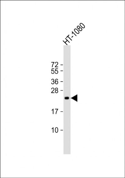
Sample:
Lane 1: Human HT-1080 cell lysates
Primary: Anti- TIMP-2 (SLM5164M) at 1/2000 dilution
Secondary: IRDye800CW Goat Anti-Mouse IgG at 1/20000 dilution
Predicted band size: 24 kD
Observed band size: 24 kD
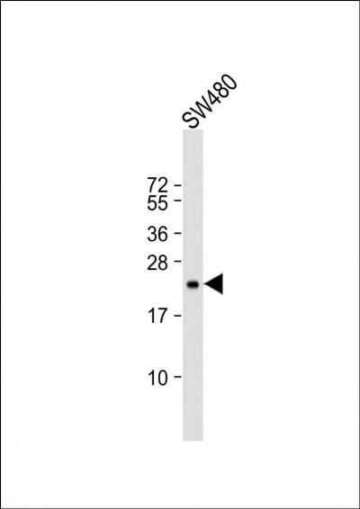
Sample:
Lane 1: Human SW96 cell lysates
Primary: Anti- TIMP-2 (SLM5164M) at 1/500 dilution
Secondary: IRDye800CW Goat Anti-Mouse IgG at 1/20000 dilution
Predicted band size: 24 kD
Observed band size: 24 kD
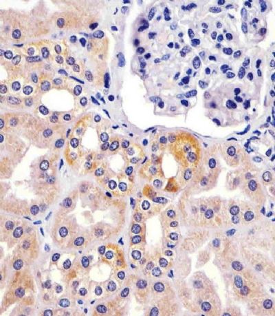
Paraformaldehyde-fixed, paraffin embedded (human kidney sections); Antigen retrieval by boiling in sodium citrate buffer (pH6.0) for 15min; Block endogenous peroxidase by 3% hydrogen peroxide for 20 minutes; Blocking buffer (normal goat serum) at 37°C for 30min; Antibody incubation with (TIMP-2) Monoclonal Antibody, Unconjugated (SLM5164M) at 1:25 overnight at 4°C, followed by operating according to SP Kit(Mouse)(sp-0024) instructionsand DAB staining.
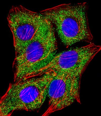
A549 cell; 4% Paraformaldehyde-fixed; Triton X-100 at room temperature for 20 min; Blocking buffer (normal goat serum, SLC0005) at 37°C for 20 min; Antibody incubation with (TIMP-2) monoclonal Antibody, Unconjugated (SLM5164M) 1:25, 90 minutes at 37°C; followed by a conjugated Goat Anti-Mouse IgG antibody at 37°C for 90 minutes, DAPI (blue, C02-04002) was used to stain the cell nuclei.
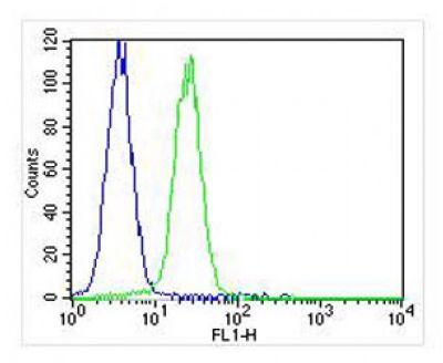
Blank control:K562.
Primary Antibody (green line): Mouse Anti-TIMP-2 antibody (SLM5164M)
Dilution:1:25;
Secondary Antibody : Goat anti-mouse IgG-AF488
Dilution:1:400.
Protocol
The cells were fixed with 2% PFA (10min at room temperature)and then permeabilized with 90% methanol for 10 min.The cells were then incubated in 2%BSA to block non-specific protein-protein interactions for 30 min at room temperature .Cells stained with Primary Antibody for 30 min at room temperature. The secondary antibody used for 40 min at room temperature. Acquisition of 20,000 events was performed.
|
|
|