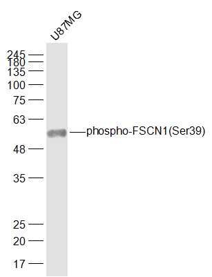
Sample:
U87MG(Human) Cell Lysate at 30 ug
Primary: Anti-phospho-FSCN1(Ser39) (SL0772R) at 1/1000 dilution
Secondary: IRDye800CW Goat Anti-Rabbit IgG at 1/20000 dilution
Predicted band size: 55 kD
Observed band size: 55 kD
Sample:
Lane 1: Mouse Cerebrum tissue lysates
Lane 2: Mouse Testis tissue lysates
Lane 3: Mouse Spleen tissue lysates
Lane 4: Rat Cerebrum tissue lysates
Lane 5: Rat Testis tissue lysates
Lane 6: Rat Spleen tissue lysates
Lane 7: Human HepG2 cell lysates
Lane 8: Human K562 cell lysates
Lane 9: Human SH-SY5Y cell lysates
Lane 10: Human U-2os cell lysates
Primary: Anti- phospho-FSCN1 (Ser39) (SL0772R) at 1/1000 dilution
Secondary: IRDye800CW Goat Anti-Rabbit IgG at 1/20000 dilution
Predicted band size: 55 kDa
Observed band size: 51 kDa
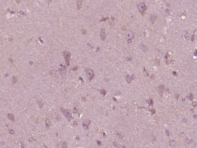
Paraformaldehyde-fixed, paraffin embedded (human brain glioma); Antigen retrieval by boiling in sodium citrate buffer (pH6.0) for 15min; Block endogenous peroxidase by 3% hydrogen peroxide for 20 minutes; Blocking buffer (normal goat serum) at 37°C for 30min; Antibody incubation with (FSCN1(Ser39)) Polyclonal Antibody, Unconjugated (SL0772R) at 1:400 overnight at 4°C, followed by operating according to SP Kit(Rabbit) (sp-0023) instructionsand DAB staining.
Tissue/cell: musle of mouse embryo; 4% Paraformaldehyde-fixed and paraffin-embedded;
Antigen retrieval: citrate buffer ( 0.01M, pH 6.0 ), Boiling bathing for 15min; Block endogenous peroxidase by 3% Hydrogen peroxide for 30min; Blocking buffer (normal goat serum,SLC0005) at 37℃ for 20 min;
Incubation: Anti-phospho-FSCN1(Ser39) Polyclonal Antibody, Unconjugated(SL0772R) 1:200, overnight at 4°C, followed by conjugation to the secondary antibody(SP-0023) and DAB(SLC0010) staining
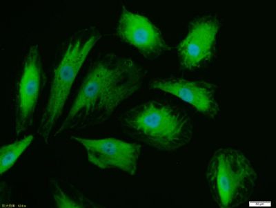
A549 cell; 4% Paraformaldehyde-fixed; Triton X-100 at room temperature for 20 min; Blocking buffer (normal goat serum, SLC0005) at 37°C for 20 min; Antibody incubation with (phospho-FSCN1 (Ser39)) polyclonal Antibody, Unconjugated (SL0772R) 1:100, 90 minutes at 37°C; followed by a conjugated Goat Anti-Rabbit IgG antibody at 37°C for 90 minutes, DAPI (blue, C02-04002) was used to stain the cell nuclei.
Blank control: U937(blue)
Isotype Control Antibody: Rabbit IgG(orange) ;
Secondary Antibody: Goat anti-rabbit IgG-FITC(white blue),
Dilution: 1:100 in 1 X PBS containing 0.5% BSA ;
Primary Antibody Dilution: 3μl in 100 μL1X PBS containing 0.5% BSA(green).
|