This gene encodes a member of the WD repeat protein family. WD repeats are minimally conserved regions of approximately 40 amino acids typically bracketed by gly-his and trp-asp (GH-WD), which may facilitate formation of heterotrimeric or multiprotein complexes. Members of this family are involved in a variety of cellular processes, including cell cycle progression, signal transduction, apoptosis, and gene regulation. Two transcript variants encoding two different isoforms have been found for this gene. [provided by RefSeq].
Function:
May be involved in MAPK pathways.
Subunit:
Interacts with DDB1-CUL4A/B E3 ligase complexes.
Subcellular Location:
Cytoplasm.
Tissue Specificity:
Broadly expressed, with highest levels in heart and skeletal muscle.
Similarity:
Contains 1 CTLH domain.
Contains 1 LisH domain.
Contains 6 WD repeats.
SWISS:
Q9H7D7
Gene ID:
80232
Database links:
Entrez Gene: 80232 Human
Entrez Gene: 226757 Mouse
Entrez Gene: 498301 Rat
SwissProt: Q9H7D7 Human
SwissProt: Q8C6G8 Mouse
Unigene: 497873 Human
Unigene: 289082 Mouse
Unigene: 23078 Rat
WDR26可能具有通过MAPK信号途径调节细胞增殖的功能。
| Picture |
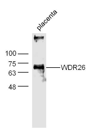
Sample:
Placenta (Mouse) Lysate at 40 ug
Primary: Anti-WDR26 (SL0932R) at 1/300 dilution
Secondary: IRDye800CW Goat Anti-Rabbit IgG at 1/20000 dilution
Predicted band size: 72 kD
Observed band size: 70 kD
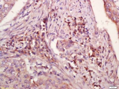
Tissue/cell: human laryngocarcinoma; 4% Paraformaldehyde-fixed and paraffin-embedded;
Antigen retrieval: citrate buffer ( 0.01M, pH 6.0 ), Boiling bathing for 15min; Block endogenous peroxidase by 3% Hydrogen peroxide for 30min; Blocking buffer (normal goat serum,SLC0005) at 37℃ for 20 min;
Incubation: Anti-WDR26 Polyclonal Antibody, Unconjugated(SL0932R) 1:200, overnight at 4°C, followed by conjugation to the secondary antibody(SP-0023) and DAB(SLC0010) staining
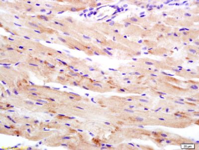
Tissue/cell: rat heart tissue; 4% Paraformaldehyde-fixed and paraffin-embedded;
Antigen retrieval: citrate buffer ( 0.01M, pH 6.0 ), Boiling bathing for 15min; Block endogenous peroxidase by 3% Hydrogen peroxide for 30min; Blocking buffer (normal goat serum,SLC0005) at 37℃ for 20 min;
Incubation: Anti-WDR26 Polyclonal Antibody, Unconjugated(SL0932R) 1:200, overnight at 4°C, followed by conjugation to the secondary antibody(SP-0023) and DAB(SLC0010) staining
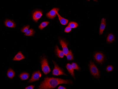
Tissue/cell: MCF7; 4% Paraformaldehyde-fixed; Triton X-100 at room temperature for 20 min; Blocking buffer (normal goat serum, SLC0005) at 37°C for 20 min; Antibody incubation with (WDR26) Polyclonal Antibody, Unconjugated (SL0932R) 1:200, 90 minutes at 37°C; followed by a conjugated Goat Anti-Rabbit IgG antibody (SL0295G-FITC) at 37°C for 90 minutes, DAPI (blue, C02-04002) was used to stain the cell nuclei.
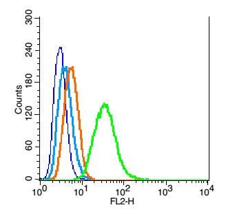
Blank control: RSC96(blue), the cells were fixed with 2% paraformaldehyde (10 min) and then permeabilized with ice-cold 90% methanol for 30 min on ice.
Isotype Control Antibody: Rabbit IgG(orange) ;
Secondary Antibody: Goat anti-rabbit IgG-PE(white blue),
Dilution: 1:200 in 1 X PBS containing 0.5% BSA ;
Primary Antibody Dilution: 1μg in 100 μL1X PBS containing 0.5% BSA(green).
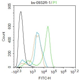
Blank control:THP-1.
Primary Antibody (green line): Rabbit Anti-WDR26 antibody (SL0932R)
Dilution: 1ug/Test;
Secondary Antibody : Goat anti-rabbit IgG-FITC
Dilution: 0.5ug/Test.
Protocol
The cells were fixed with 4% PFA (10min at room temperature)and then permeabilized with 90% ice-cold methanol for 20 min at -20℃.The cells were then incubated in 5%BSA to block non-specific protein-protein interactions for 30 min at room temperature .Cells stained with Primary Antibody for 30 min at room temperature. The secondary antibody used for 40 min at room temperature. Acquisition of 20,000 events was performed.
|
|
|