Aquaporin 1 is a 28kD integral membrane protein which was originally identified in red blood cells and renal proximal tubules. AQP1 is also expressed by the choroid plexus and various other tissues. It forms a water-specific channel that provides the plasma membranes of red cells and kidney proximal tubules with high permeability to water, thereby permitting water to move in the direction of an osmotic gradient.
Function:
Forms a water-specific channel that provides the plasmamembranes of red cells and kidney proximal tubules with highpermeability to water, thereby permitting water to move in thedirection of an osmotic gradient.
Subunit:
Homotetramer. Interacts with EPHB2; involved in endolymphproduction in the inner ear (By similarity).
Subcellular Location:
Membrane; Multi-pass membrane protein.
Tissue Specificity:
Expressed in a number of tissues including erythrocytes, renal tubules, retinal pigment epithelium, heart, lung, skeletal muscle, kidney and pancreas. Weakly expressed in brain, placenta and liver.
Similarity:
Belongs to the MIP/aquaporin (TC 1.A.8) family.
SWISS:
P29972
Gene ID:
358
Database links:
Entrez Gene: 358 Human
Entrez Gene: 11826 Mouse
Entrez Gene: 2548 Rat
Entrez Gene: 442999 Sheep
Entrez Gene: 282653 Cow
Entrez Gene: 403732 Dog
Omim: 107776 Human
SwissProt: P47865 Cow
SwissProt: Q9N2J4 Dog
SwissProt: P29972 Human
SwissProt: Q02013 Mouse
SwissProt: P29975 Rat
SwissProt: P51281 Sheep
Unigene: 76152 Human
Unigene: 18625 Mouse
Unigene: 1618 Rat
通道蛋白(Channel Protein)
| Picture |
Sample:
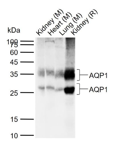
Lane 1: Mouse Kidney tissue lysates
Lane 2: Mouse Heart tissue lysates
Lane 3: Mouse Lung tissue lysates
Lane 4: Rat Kidney tissue lysates
Primary: Anti-AQP1 (SL1506R) at 1/1000 dilution
Secondary: IRDye800CW Goat Anti-Rabbit IgG at 1/20000 dilution
Predicted band size: 28 kDa
Observed band size: 35,28 kDa
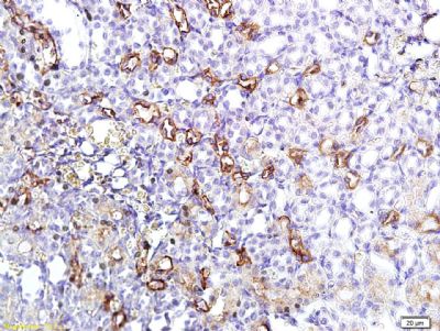
Tissue/cell: rat kidney tissue; 4% Paraformaldehyde-fixed and paraffin-embedded;
Antigen retrieval: citrate buffer ( 0.01M, pH 6.0 ), Boiling bathing for 15min; Block endogenous peroxidase by 3% Hydrogen peroxide for 30min; Blocking buffer (normal goat serum,SLC0005) at 37℃ for 20 min;
Incubation: Anti-AQP-1 Polyclonal Antibody, Unconjugated(SL1506R) 1:400, overnight at 4°C, followed by conjugation to the secondary antibody(SP-0023) and DAB(SLC0010) staining
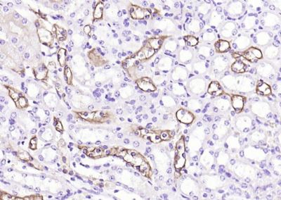
Paraformaldehyde-fixed, paraffin embedded (mouse kidney); Antigen retrieval by boiling in sodium citrate buffer (pH6.0) for 15min; Block endogenous peroxidase by 3% hydrogen peroxide for 20 minutes; Blocking buffer (normal goat serum) at 37°C for 30min; Antibody incubation with (AQP1) Polyclonal Antibody, Unconjugated (SL1506R) at 1:200 overnight at 4°C, followed by operating according to SP Kit(Rabbit) (sp-0023) instructionsand DAB staining.
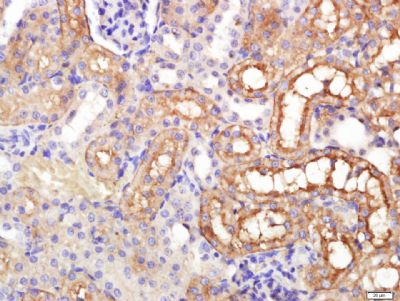
Paraformaldehyde-fixed, paraffin embedded (rat kidney); Antigen retrieval by boiling in sodium citrate buffer (pH6.0) for 15min; Block endogenous peroxidase by 3% hydrogen peroxide for 20 minutes; Blocking buffer (normal goat serum) at 37°C for 30min; Antibody incubation with (AQP1) Polyclonal Antibody, Unconjugated (SL1506R) at 1:200 overnight at 4°C, followed by operating according to SP Kit(Rabbit) (sp-0023) instructionsand DAB staining.
Paraformaldehyde-fixed, paraffin embedded (rat kidney); Antigen retrieval by boiling in sodium citrate buffer (pH6.0) for 15min; Block endogenous peroxidase by 3% hydrogen peroxide for 20 minutes; Blocking buffer (normal goat serum) at 37°C for 30min; Antibody incubation with (AQP1) Polyclonal Antibody, Unconjugated (SL1506R) at 1:200 overnight at 4°C, followed by a conjugated secondary (sp-0023) for 20 minutes and DAB staining.
Blank control: 293T (blue).
Primary Antibody:Rabbit Anti-AQP1 antibody(SL1506R), Dilution: 1μg in 100μL 1X PBS containing 0.5% BSA;
Isotype Control Antibody: Rabbit IgG(orange) ,used under the same conditions );
Secondary Antibody: Goat anti-rabbit IgG-PE(white blue), Dilution: 1:200 in 1 X PBS containing 0.5% BSA.
Protocol
The cells were fixed with 2% paraformaldehyde (10 min). Primary antibody (SL1506R, 1μg /1x10^6 cells) were incubated for 30 min on the ice, followed by 1 X PBS containing 0.5% BSA + 10% goat serum (15 min) to block non-specific protein-protein interactions. Then the Goat Anti-rabbit IgG/PE antibody was added into the blocking buffer mentioned above to react with the primary antibody at 1/200 dilution for 30 min on ice. Acquisition of 20,000 events was performed.
|
|
|