Protein tyrosine phosphatases (PTPases) SHP1 and SHP2 are critical regulators in the intracellular signaling pathways that result in cell responses such as mitosis, differentiation, migration, survival, transformation or death. SHP2 is a signal transducer for several receptor tyrosine kinases and cytokine receptors. A novel SHP2 associated glycoprotein was recently cloned from human, rat, mouse and cattle by several labs and was designated SIRPa (1),SHPS1 , MyD1, BIT and p84. SIRPa is a new gene family containing at least fifteen members. SIRPa is a substrate of many activated tyrosine kinases such as insulin receptor, EGFR, PDGFR and src, and a specific docking protein for SHP2. SIRPa has regulatory effects on cellular responses induced by serum, growth factors, insulin, oncogenes, growth hormones and cell adhesion and plays a general role in different physiological and pathological processes.
Function:
Immunoglobulin-like cell surface receptor for CD47. Acts as docking protein and induces translocation of PTPN6, PTPN11 and other binding partners from the cytosol to the plasma membrane. Supports adhesion of cerebellar neurons, neurite outgrowth and glial cell attachment. May play a key role in intracellular signaling during synaptogenesis and in synaptic function (By similarity). Involved in the negative regulation of receptor tyrosine kinase-coupled cellular responses induced by cell adhesion, growth factors or insulin. Mediates negative regulation of phagocytosis, mast cell activation and dendritic cell activation. CD47 binding prevents maturation of immature dendritic cells and inhibits cytokine production by mature dendritic cells.
Subunit:
Binds PTPN11 when tyrosine-phosphorylated, except in macrophages, where it primarily binds PTPN6. Binds GRB2 in vitro. Binds FGR (By similarity). Binds JAK2 irrespective of its phosphorylation status and forms a stable complex. Binds SCAP1 and/or SCAP2. The resulting complex recruits FYB. Binds PTK2B.
Subcellular Location:
Membrane; Single-pass type I membraneprotein.
Tissue Specificity:
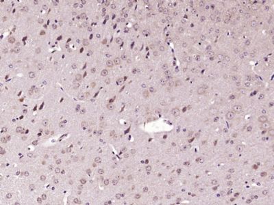
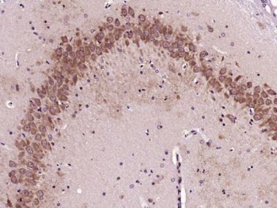
Ubiquitous. Highly expressed in brain. Detected on myeloid cells, but not T-cells. Detected at lower levels in heart, placenta, lung, testis, ovary, colon, liver, small intestine, prostate, spleen, kidney, skeletal muscle and pancreas.
Post-translational modifications:
N-glycosylated.
Phosphorylated on tyrosine residues in response to stimulation with EGF, growth hormone, insulin and PDGF.
Dephosphorylated by PTPN11.
Similarity:
Contains 2 Ig-like C1-type (immunoglobulin-like) domains.
Contains 1 Ig-like SLVtype (immunoglobulin-like) domain.
SWISS:
P78324
Gene ID:
140885
Database links:
Entrez Gene: 140885 Human
Entrez Gene: 19261 Mouse
Entrez Gene: 25528 Rat
Omim: 602461 Human
SwissProt: P78324 Human
SwissProt: P97797 Mouse
SwissProt: P97710 Rat
Unigene: 581021 Human
Unigene: 1682 Mouse
Unigene: 53971 Rat
| Picture |
Sample:
Cerebrum (Mouse) Lysate at 40 ug
Primary: Anti-SIRP Alpha (SL2708R) at 1/1000 dilution
Secondary: IRDye800CW Goat Anti-Rabbit IgG at 1/20000 dilution
Predicted band size: 56 kD
Observed band size: 56 kD
Sample:
Spleen(Mouse) Lysate at 30 ug
Bones (Mouse) Lysate at 30 ug
Primary: Anti- SIRP Alpha (SL2708) at 1/300 dilution
Secondary: IRDye800CW Goat Anti-Rabbit IgG at 1/20000 dilution
Predicted band size: 56 kD
Observed band size: 53 kD
Sample:
Lane 1: Mouse Brain Lysates
Lane 2: Mouse Cerebellum Lysates
Lane 3: Mouse Spleen Lysates
Lane 4: Mouse Placenta Lysates
Lane 5: Rat Brain Lysates
Lane 6: Rat Cerebellum Lysates
Primary: Anti-SIRP Alpha(SL2708R) at 1/1000 dilution
Secondary: IRDye800CW Goat Anti-Rabbit IgG at 1/20000 dilution
Predicted band size: 56kDa
Observed band size: 56kDa
Sample:
Raw264.7(Mouse) Cell Lysate at 30 ug
Primary: Anti-SIRP Alpha (SL2708R) at 1/1000 dilution
Secondary: IRDye800CW Goat Anti-Rabbit IgG at 1/20000 dilution
Predicted band size: 56 kD
Observed band size: 56 kD
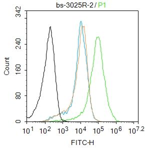
Blank control(blue): U937 (fixed with 2% paraformaldehyde (10 min)).
Primary Antibody:Rabbit Anti- SIRP antibody(SL2708R), Dilution: 1μg in 100 μL 1X PBS containing 0.5% BSA;
Isotype Control Antibody: Rabbit IgG(orange) ,used under the same conditions );
Secondary Antibody: Goat anti-rabbit IgG-PE(white blue), Dilution: 1:200 in 1 X PBS containing 0.5% BSA.
|
|
|