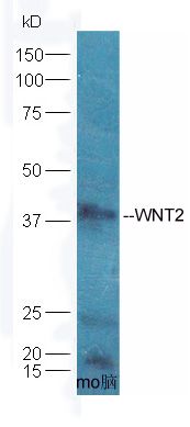
Sample: Brain (Mouse) Lysate at 40 ug
Primary: Anti- WNT2 (SL6133R) at 1/300 dilution
Secondary: HRP conjugated Goat-Anti-rabbit IgG (SL0295G-HRP) at 1/5000 dilution
Predicted band size: 37 kD
Observed band size: 38 kD
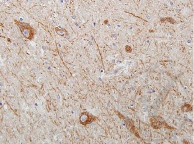
Images provided the Independent Validation Program (badge number 029629)Formalin-fixed and paraffin embedded human brain tissue with Parkinson's morphology labeled with Rabbit Anti-WNT2 Polyclonal Antibody (SL6133R) at 1:250 overnight at 4 °C followed by conjugation to secondary antibody.
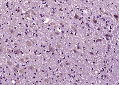
Paraformaldehyde-fixed, paraffin embedded (mouse cerebellum); Antigen retrieval by boiling in sodium citrate buffer (pH6.0) for 15min; Block endogenous peroxidase by 3% hydrogen peroxide for 20 minutes; Blocking buffer (normal goat serum) at 37°C for 30min; Antibody incubation with (WNT2) Polyclonal Antibody, Unconjugated (SL6133R) at 1:200 overnight at 4°C, followed by operating according to SP Kit(Rabbit) (sp-0023) instructionsand DAB staining.
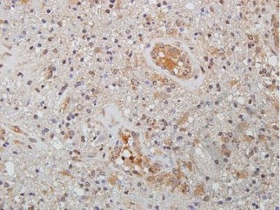
Images provided the Independent Validation Program (badge number 029629)Formalin-fixed and paraffin embedded human brain glioma tissue labeled with Rabbit Anti-WNT2 Polyclonal Antibody (SL6133R) at 1:250 overnight at 4 °C followed by conjugation to secondary antibody.
|