Home > Product > Antibody > Rabbit Anti-EphA7 antibody
Cek 11; Developmental kinase 1; EBK; EHK-3; EHK3; EK11; Embryonic brain kinase; Eph homology kinase 3; Eph receptor A7; EPH-like kinase 11; EphA7; EPHA7_HUMAN; Ephrin receptor Eph A7; Ephrin type A receptor 7; Ephrin type-A receptor 7; hEK11; MDK 1; Recep
Cat:
SL7034R
Species Reactivity:
Human,Mouse,Rat,(predicted: Chicken,Dog,Cow,Horse,Rabbit,)
Immunogen:
KLH conjugated synthetic peptide derived from human EphA7:181-280/998<Extracellular>
Format:
Liquid
Storage instructions:
Shipped at 4℃. Store at -20 °C for one year. Avoid repeated freeze/thaw cycles.
Concentration:
1mg/ml
Clonality:
Polyclonal
Isotype:
IgG
Applications:
WB=1:500-2000ELISA=1:5000-10000IHC-P=1:100-500IHC-F=1:100-500IF=1:100-500(Paraffin sections need to do antigen repair)not yet tested in other applications.optimal dilutions/concentrations should be determined by the end user.
Host:
Rabbit
Product Overview:
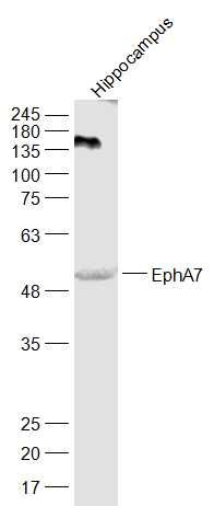
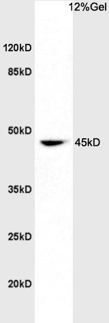
Sample: Cerebrum (Rat) Lysate at 40 ugPrimary: Anti-EphA7 (SL7034R) at 1/1000 dilutionSecondary: IRDye800CW Goat Anti-Rabbit IgG at 1/20000 dilutionPredicted band size: 50/107 kDObserved band size: 52 kDProtein: rat brain lysates, 30ug; Primary: Anti-EphA7 (SL7034R) at 1:200; Secondary: HRP conjugated Goat Anti-Rabbit IgG(SL0295G-HRP) at 1: 3000; ECL excitated the fluorescence; Predicted band size : 107kDObserved band size : 45kDSample: Hippocampus (Mouse) Lysate at 40 ugPrimary: Anti-EphA7 (SL7034R) at 1/1000 dilutionSecondary: IRDye800CW Goat Anti-Rabbit IgG at 1/20000 dilutionPredicted band size: 50/107 kDObserved band size: 52 kDTissue/cell: rat brain tissue(hippocampus); 4% Paraformaldehyde-fixed and paraffin-embedded; Antigen retrieval: citrate buffer ( 0.01M, pH 6.0 ), Boiling bathing for 15min; Block endogenous peroxidase by 3% Hydrogen peroxide for 30min; Blocking buffer (normal goat serum,SLC0005) at 37℃ for 20 min; Incubation: Anti-EphA7 Polyclonal Antibody, Unconjugated(SL7034R) 1:200, overnight at 4°C, followed by conjugation to the secondary antibody(SP-0023) and DAB(SLC0010) staining
More
Unit:
Price: $
Product PDFs
Datasheet:


Protein kinases are enzymes that transfer a phosphate group from a phosphate donor, generally the g phosphate of ATP, onto an acceptor amino acid in a substrate protein. By this basic mechanism, protein kinases mediate most of the signal transduction in eukaryotic cells, regulating cellular metabolism, transcription, cell cycle progression, cytoskeletal rearrangement and cell movement, apoptosis, and differentiation. With more than 500 gene products, the protein kinase family is one of the largest families of proteins in eukaryotes. The family has been classified in 8 major groups based on sequence comparison of their tyrosine (PTK) or serine/threonine (STK) kinase catalytic domains. The tyrosine kinase (TK) group is mainly involved in the regulation of cell-cell interactions such as differentiation, adhesion, motility and death. There are currently about 90 TK genes sequenced, 58 are of receptor protein TK (e.g. EGFR, EPH, FGFR, PDGFR, TRK, and VEGFR families), and 32 of cytosolic TK (e.g. ABL, FAK, JAK, and SRC families).
Receptor for members of the ephrin-A family. Binds to ephrin-A1, ephrin-A2, ephrin-A3, ephrin-A4 and ephrin-A5.

Function:
’Receptor tyrosine kinase which binds promiscuouslyGPI-anchored ephrin-A family ligands residing on adjacent cells,leading to contact-dependent bidirectional signaling intoneighboring cells. The signaling pathway downstream of the receptoris referred to as forward signaling while the signaling pathwaydownstream of the ephrin ligand is referred to as reversesignaling. Among GPI-anchored ephrin-A ligands, EFNA5 is acognate/functional ligand for EPHA7 and their interaction regulatesbrain development modulating cell-cell adhesion and repulsion. Hasa repellent activity on axons and is for instance involved in theguidance of corticothalamic axons and in the proper topographicmapping of retinal axons to the colliculus. May also regulate braindevelopment through a caspase(CASP3)-dependent proapoptoticactivity. Forward signaling may result in activation of componentsof the ERK signaling pathway including MAP2K1, MAP2K2, MAPK1 ANDMAPK3 which are phosphorylated upon activation of EPHA7. Isoform 4which lacks the kinase domain may regulate isoform 1 adhesiveproperties.

Subunit:
Heterotetramer upon binding of the ligand. The heterotetramer is composed of an ephrin dimer and a receptor dimer. Oligomerization is probably required to induce biological responses (By similarity). Interacts (via PDZ-binding motif) with GRIP1 and PICK1 (via PDZ domain).

Subcellular Location:
Cell membrane (Probable); Single-pass type I membrane protein (Probable).

Tissue Specificity:
Widely expressed in embryo. In adult, expression restricted to hippocampus, testis and spleen.

Post-translational modifications:
Phosphorylated. Isoform 4 inhibits isoform 1 phosphorylationand may regulate its function in adhesion.

Similarity:
Belongs to the protein kinase superfamily. Tyr protein kinase family. Ephrin receptor subfamily.
Contains 1 Eph LBD (Eph ligand-binding) domain.
Contains 2 fibronectin type-III domains.
Contains 1 protein kinase domain.
Contains 1 SAM (sterile alpha motif) domain.

SWISS:
Q15375

Gene ID:
2045

Database links:

Entrez Gene: 2045 Human

Entrez Gene: 13841 Mouse

Entrez Gene: 171287 Rat

Omim: 602190 Human

SwissProt: Q15375 Human

SwissProt: Q61772 Mouse

SwissProt: P54759 Rat

Unigene: 73962 Human

Unigene: 257266 Mouse

Unigene: 10181 Rat


Picture

Sample:
Cerebrum (Rat) Lysate at 40 ug
Primary: Anti-EphA7 (SL7034R) at 1/1000 dilution
Secondary: IRDye800CW Goat Anti-Rabbit IgG at 1/20000 dilution
Predicted band size: 50/107 kD
Observed band size: 52 kD
Protein: rat brain lysates, 30ug;
Primary: Anti-EphA7 (SL7034R) at 1:200;
Secondary: HRP conjugated Goat Anti-Rabbit IgG(SL0295G-HRP) at 1: 3000;
ECL excitated the fluorescence;
Predicted band size : 107kD
Observed band size : 45kD
Sample:
Hippocampus (Mouse) Lysate at 40 ug
Primary: Anti-EphA7 (SL7034R) at 1/1000 dilution
Secondary: IRDye800CW Goat Anti-Rabbit IgG at 1/20000 dilution
Predicted band size: 50/107 kD
Observed band size: 52 kD
Tissue/cell: rat brain tissue(hippocampus); 4% Paraformaldehyde-fixed and paraffin-embedded;
Antigen retrieval: citrate buffer ( 0.01M, pH 6.0 ), Boiling bathing for 15min; Block endogenous peroxidase by 3% Hydrogen peroxide for 30min; Blocking buffer (normal goat serum,SLC0005) at 37℃ for 20 min;
Incubation: Anti-EphA7 Polyclonal Antibody, Unconjugated(SL7034R) 1:200, overnight at 4°C, followed by conjugation to the secondary antibody(SP-0023) and DAB(SLC0010) staining
Product Feedback Wall
Message :
Your Email :
Copyright © 2007-2018 Sunlong Medical All Rights Reserved.