Home > Product > Antibody > Rabbit Anti-ENC1 antibody
CCL 28; CCL28; Ectoderm neural cortex protein 1; Ectoderm-neural cortex protein 1; Ectodermal neural cortex (with BTB like domain); ENC 1; ENC-1; ENC1; ENC1_HUMAN; FLJ39259; Kelch like 35; Kelch like 37; Kelch-like protein 37; KLHL 35; KLHL35; KLHL37; NRP
Cat:
SL8043R
Species Reactivity:
Mouse,Rat,(predicted: Human,Cow,Horse,Rabbit,Sheep,)
Immunogen:
KLH conjugated synthetic peptide derived from human ENC1:241-300/589
Format:
Liquid
Storage instructions:
Shipped at 4℃. Store at -20 °C for one year. Avoid repeated freeze/thaw cycles.
Concentration:
1mg/ml
Clonality:
Polyclonal
Isotype:
IgG
Applications:
WB=1:500-2000ELISA=1:5000-10000IHC-P=1:100-500IHC-F=1:100-500Flow-Cyt=0.2ug/testIF=1:100-500(Paraffin sections need to do antigen repair)not yet tested in other applications.optimal dilutions/concentrations should be determined by the end user.
Host:
Rabbit
Product Overview:
Paraformaldehyde-fixed, paraffin embedded (Mouse brain); Antigen retrieval by boiling in sodium citrate buffer (pH6.0) for 15min; Block endogenous peroxidase by 3% hydrogen peroxide for 20 minutes; Blocking buffer (normal goat serum) at 37°C for 30min; Antibody incubation with (ENC1) Polyclonal Antibody, Unconjugated (SL8043R) at 1:400 overnight at 4°C, followed by operating according to SP Kit(Rabbit) (sp-0023) instructionsand DAB staining. Paraformaldehyde-fixed, paraffin embedded (Rat brain); Antigen retrieval by boiling in sodium citrate buffer (pH6.0) for 15min; Block endogenous peroxidase by 3% hydrogen peroxide for 20 minutes; Blocking buffer (normal goat serum) at 37°C for 30min; Antibody incubation with (ENC1) Polyclonal Antibody, Unconjugated (SL8043R) at 1:400 overnight at 4°C, followed by operating according to SP Kit(Rabbit) (sp-0023) instructionsand DAB staining.Blank control: A549. Primary Antibody (green line): Rabbit Anti-ENC1 antibody (SL8043R) Dilution: 0.2μg /10^6 cells; Isotype Control Antibody (orange line): Rabbit IgG . Secondary Antibody : Goat anti-rabbit IgG-PEDilution: 1μg /test. ProtocolThe cells were fixed with 4% PFA (10min at room temperature)and then permeabilized with 90% ice-cold methanol for 20 min at-20℃. The cells were then incubated in 5%BSA to block non-specific protein-protein interactions for 30 min at at room temperature .Cells stained with Primary Antibody for 30 min at room temperature. The secondary antibody used for 40 min at room temperature. Acquisition of 20,000 events was performed.Blank control: A549. Primary Antibody (green line): Rabbit Anti-ENC1 antibody (SL8043R) Dilution: 1μg /10^6 cells; Isotype Control Antibody (orange line): Rabbit IgG . Secondary Antibody : Goat anti-rabbit IgG-PEDilution: 3μg /test. ProtocolThe cells were fixed with 4% PFA (10min at room temperature)and then permeabilized with 90% ice-cold methanol for 20 min at-20℃. The cells were then incubated in 5% BSA to block non-specific protein-protein interactions for 30 min at at room temperature .Cells stained with Primary Antibody for 30 min at room temperature. The secondary antibody used for 40 min at room temperature. Acquisition of 20,000 events was performed.
More
Unit:
Price: $
Product PDFs
Datasheet:


Actin-binding protein involved in the regulation of neuronal process formation and in differentiation of neural crest cells. May be down-regulated in neuroblastoma tumors. Substrate-specific adapter of an E3 ubiquitin-protein ligase complex which mediates the ubiquitination and subsequent proteasomal degradation of target proteins.
Tissue specificity: Detected in fetal brain tissue, moderate expression in fetal heart, lung and kidney. Highly expressed in adult brain, particularly high in the hippocampus and amygdala, and spinal chord. Detectable in adult pancreas.

Function:
Actin-binding protein involved in the regulation of neuronal process formation and in differentiation of neural crest cells. May be down-regulated in neuroblastoma tumors. Substrate-specific adapter of an E3 ubiquitin-protein ligase complex which mediates the ubiquitination and subsequent proteasomal degradation of target proteins.

Subunit:
Binds to RB1. Hypophosphorylated RB1 associates with ENC1 during neuronal differentiation, while hyperphosphorylated RB1 associates with ENC1 in undifferentiating cells. Part of a complex that contains CUL3, RBX1 and ENC1.

Subcellular Location:
Nucleus matrix. Cytoplasm, cytoskeleton.

Tissue Specificity:
Detected in fetal brain tissue, moderate expression in fetal heart, lung and kidney. Highly expressed in adult brain, particularly high in the hippocampus and amygdala, and spinal chord. Detectable in adult pancreas.

Post-translational modifications:
Ubiquitinated and probably targeted for proteasome-independent degradation.

Similarity:
Contains 1 BTB (POZ) domain.
Contains 6 Kelch repeats.

SWISS:
O14682

Gene ID:
8507

Database links:

Entrez Gene: 8507 Human

Entrez Gene: 2763 Mouse

Entrez Gene: 294674 Rat

Omim: 605173 Human

SwissProt: O14682 Human

SwissProt: O35709 Mouse

Unigene: 104925 Human

Unigene: 241073 Mouse


Picture

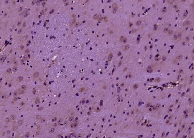
Paraformaldehyde-fixed, paraffin embedded (Mouse brain); Antigen retrieval by boiling in sodium citrate buffer (pH6.0) for 15min; Block endogenous peroxidase by 3% hydrogen peroxide for 20 minutes; Blocking buffer (normal goat serum) at 37°C for 30min; Antibody incubation with (ENC1) Polyclonal Antibody, Unconjugated (SL8043R) at 1:400 overnight at 4°C, followed by operating according to SP Kit(Rabbit) (sp-0023) instructionsand DAB staining.
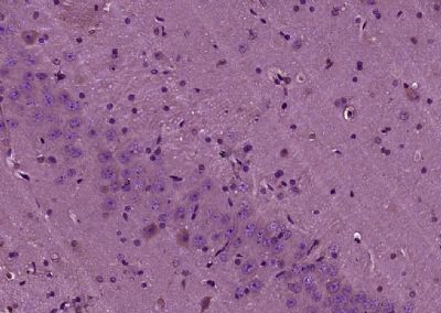
Paraformaldehyde-fixed, paraffin embedded (Rat brain); Antigen retrieval by boiling in sodium citrate buffer (pH6.0) for 15min; Block endogenous peroxidase by 3% hydrogen peroxide for 20 minutes; Blocking buffer (normal goat serum) at 37°C for 30min; Antibody incubation with (ENC1) Polyclonal Antibody, Unconjugated (SL8043R) at 1:400 overnight at 4°C, followed by operating according to SP Kit(Rabbit) (sp-0023) instructionsand DAB staining.
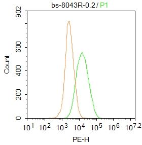
Blank control: A549.
Primary Antibody (green line): Rabbit Anti-ENC1 antibody (SL8043R)
Dilution: 0.2μg /10^6 cells;
Isotype Control Antibody (orange line): Rabbit IgG .
Secondary Antibody : Goat anti-rabbit IgG-PE
Dilution: 1μg /test.
Protocol
The cells were fixed with 4% PFA (10min at room temperature)and then permeabilized with 90% ice-cold methanol for 20 min at-20℃. The cells were then incubated in 5%BSA to block non-specific protein-protein interactions for 30 min at at room temperature .Cells stained with Primary Antibody for 30 min at room temperature. The secondary antibody used for 40 min at room temperature. Acquisition of 20,000 events was performed.
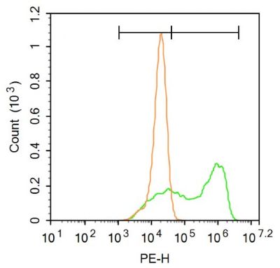
Blank control: A549.
Primary Antibody (green line): Rabbit Anti-ENC1 antibody (SL8043R)
Dilution: 1μg /10^6 cells;
Isotype Control Antibody (orange line): Rabbit IgG .
Secondary Antibody : Goat anti-rabbit IgG-PE
Dilution: 3μg /test.
Protocol
The cells were fixed with 4% PFA (10min at room temperature)and then permeabilized with 90% ice-cold methanol for 20 min at-20℃. The cells were then incubated in 5% BSA to block non-specific protein-protein interactions for 30 min at at room temperature .Cells stained with Primary Antibody for 30 min at room temperature. The secondary antibody used for 40 min at room temperature. Acquisition of 20,000 events was performed.
Product Feedback Wall
Message :
Your Email :
Copyright © 2007-2018 Sunlong Medical All Rights Reserved.