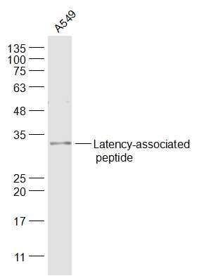
Sample:
A549(Human) Cell Lysate at 30 ug
Primary: Anti-Latency-associated peptide (SL4908R) at 1/1000 dilution
Secondary: IRDye800CW Goat Anti-Rabbit IgG at 1/20000 dilution
Predicted band size: 30/44 kD
Observed band size: 30 kD
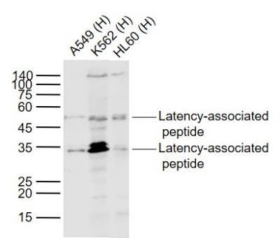
Sample:
Lane 1: A549 (Human) Cell Lysate at 30 ug
Lane 2: K562 (Human) Cell Lysate at 30 ug
Lane 3: HL60 (Human) Cell Lysate at 30 ug
Primary:
Anti-Latency-associated peptide (SL4908R) at 1/1000 dilution
Secondary: IRDye800CW Goat Anti-Rabbit IgG at 1/20000 dilution
Predicted band size: 50/35 kD
Observed band size: 50/33 kD
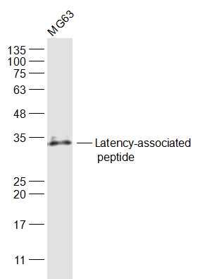
Sample:
MG63(Human) Cell Lysate at 30 ug
Primary: Anti-Latency-associated peptide (SL4908R) at 1/1000 dilution
Secondary: IRDye800CW Goat Anti-Rabbit IgG at 1/20000 dilution
Predicted band size: 30/44 kD
Observed band size: 30 kD
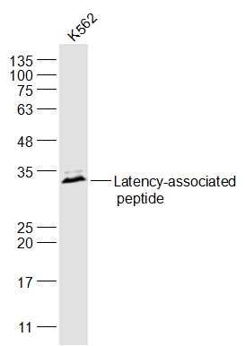
Sample:
K562(Human) Cell Lysate at 30 ug
Primary: Anti-Latency-associated peptide (SL4908R) at 1/1000 dilution
Secondary: IRDye800CW Goat Anti-Rabbit IgG at 1/20000 dilution
Predicted band size: 30/44 kD
Observed band size: 30 kD
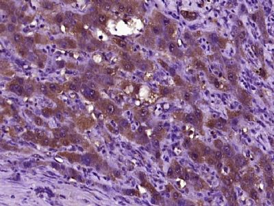
Paraformaldehyde-fixed, paraffin embedded (human liver carcinoma); Antigen retrieval by boiling in sodium citrate buffer (pH6.0) for 15min; Block endogenous peroxidase by 3% hydrogen peroxide for 20 minutes; Blocking buffer (normal goat serum) at 37°C for 30min; Antibody incubation with (TGFB1) Polyclonal Antibody, Unconjugated (SL4908R) at 1:400 overnight at 4°C, followed by operating according to SP Kit(Rabbit) (sp-0023) instructionsand DAB staining.
|