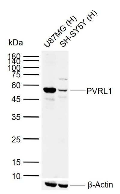
Sample:
Lane 1: Human U87MG cell lysates
Lane 2: Human SH-SY5Y cell lysates
Primary: Anti-PVRL1 (SL11126R) at 1/1000 dilution
Secondary: IRDye800CW Goat Anti-Rabbit IgG at 1/20000 dilution
Predicted band size: 54 kDa
Observed band size: 54 kDa
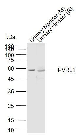
Sample:
Lane 1: Mouse Urinary bladder tissue lysates
Lane 2: Rat Urinary bladder tissue lysates
Primary: Anti-PVRL1 (SL11126R) at 1/1000 dilution
Secondary: IRDye800CW Goat Anti-Rabbit IgG at 1/20000 dilution
Predicted band size: 54 kDa
Observed band size: 57 kDa
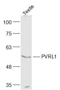
Sample:
Testis(Mouse) Lysate at 40 ug
Primary: Anti-PVRL1 (SL11126R) at 1/1000 dilution
Secondary: IRDye800CW Goat Anti-Rabbit IgG at 1/20000 dilution
Predicted band size: 54 kD
Observed band size: 54 kD
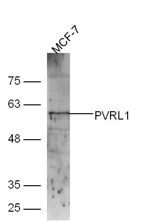
Sample:
MCF-7 Cell (Human) Lysate at 30 ug
Primary: Anti-PVRL1 (SL11126R) at 1/300 dilution
Secondary: IRDye800CW Goat Anti-Rabbit IgG at 1/20000 dilution
Predicted band size: 54 kD
Observed band size: 57 kD
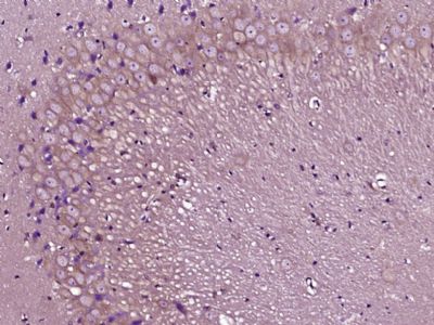
Paraformaldehyde-fixed, paraffin embedded (Rat brain); Antigen retrieval by boiling in sodium citrate buffer (pH6.0) for 15min; Block endogenous peroxidase by 3% hydrogen peroxide for 20 minutes; Blocking buffer (normal goat serum) at 37°C for 30min; Antibody incubation with (PVRL1) Polyclonal Antibody, Unconjugated (SL11126R) at 1:400 overnight at 4°C, followed by operating according to SP Kit(Rabbit) (sp-0023) instructionsand DAB staining.
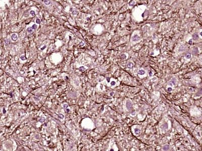
Paraformaldehyde-fixed, paraffin embedded (Rat brain); Antigen retrieval by boiling in sodium citrate buffer (pH6.0) for 15min; Block endogenous peroxidase by 3% hydrogen peroxide for 20 minutes; Blocking buffer (normal goat serum) at 37°C for 30min; Antibody incubation with (PVRL1) Polyclonal Antibody, Unconjugated (SL11126R) at 1:400 overnight at 4°C, followed by operating according to SP Kit(Rabbit) (sp-0023) instructionsand DAB staining.
|