Chromosome 9 consists of about 145 million bases and 4% of the human genome and encodes nearly 900 genes. Considered to play a role in gender determination, deletion of the distal portion of 9p can lead to development of male to female sex reversal, the phenotype of a female with a male X,Y genotype. Hereditary hemorrhagic telangiectasia, which is characterized by harmful vascular defects, is associated with the chromosome 9 gene encoding endoglin protein, ENG. Familial dysautonomia is also associated with chromosome 9 though through the gene IKBKAP. Notably, chromosome 9 encompasses the largest interferon family gene cluster. Chromosome 9 is partnered with chromosome 22 in the translocation leading to the aberrant production of BCR-ABL fusion protein often found in leukemias. The C9orf72 gene product has been provisionally designated C9orf72 pending further characterization. There are two isoforms of C9orf72 that are produced as a result of alternative splicing events.
Subcellular Location:
Cytoplasm. Nucleus. Note=Detected in the cytoplasm of neurons from post mortem brain tissue (PubMed:21944778). Detected in the nucleus in fibroblasts (PubMed:21944779).
Tissue Specificity:
Both isoforms are widely expressed, including kidney, lung, liver, heart, testis and several brain regions, such as cerebellum. Also expressed in the frontal cortex and in lymphoblasts (at protein level).
DISEASE:
Defects in C9orf72 are the cause of frontotemporal dementia and/or amyotrophic lateral sclerosis (FTDALS) [MIM:105550]. An autosomal dominant neurodegenerative disorder characterized by adult onset of frontotemporal dementia and/or amyotrophic lateral sclerosis in an affected individual. There is high intrafamilial variation. Frontotemporal dementia is characterized by frontal and temporal lobe atrophy associated with neuronal loss, gliosis, and dementia. Patients exhibit progressive changes in social, behavioral, and/or language function. Amyotrophic lateral sclerosis is characterized by the death of motor neurons in the brain, brainstem, and spinal cord, resulting in fatal paralysis. Note=Caused by a large expansion of a GGGGCC hexanucleotide within the first C9orf72 intron located between the first and the second non-coding exons. The expansion leads to the loss of transcription of one of the two transcripts encoding isoform 1 and to the formation of nuclear RNA foci.
SWISS:
Q96LT7
Gene ID:
203228
Database links:
Entrez Gene: 203228 Human
Entrez Gene: 7645 Mouse
Entrez Gene: 313155 Rat
Omim: 614260 Human
SwissProt: Q96LT7 Human
SwissProt: Q6DFW0 Mouse
SwissProt: Q66HC3 Rat
Unigene: 493639 Human
Unigene: 331544 Mouse
Unigene: 233897 Rat
| Picture |
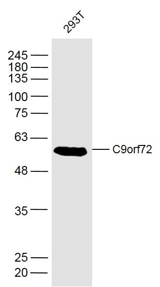
Sample:
293T(Human) Cell Lysate at 40 ug
Primary: Anti-C9orf72 (SL8595R) at 1/300 dilution
Secondary: IRDye800CW Goat Anti-Rabbit IgG at 1/20000 dilution
Predicted band size: 53 kD
Observed band size: 53 kD
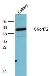
Sample:
Kidney(Rat) Lysate at 40 ug
Primary: Anti-C9orf72 (SL8595R) at 1/1000 dilution
Secondary: IRDye800CW Goat Anti-Rabbit IgG at 1/20000 dilution
Predicted band size: 53 kD
Observed band size: 53 kD
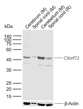
Sample:
Lane 1: Mouse Cerebrum tissue lysates
Lane 2: Mouse Spinal cord tissue lysates
Lane 3: Mouse Cerebellum tissue lysates
Lane 4: Rat Spinal cord tissue lysates
Primary: Anti- C9orf72 (SL8595R) at 1/1000 dilution
Secondary: IRDye800CW Goat Anti-Rabbit IgG at 1/20000 dilution
Predicted band size: 53 kDa
Observed band size: 50 kDa
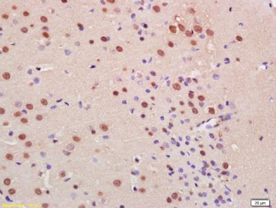
Tissue/cell: human liver carcinoma; 4% Paraformaldehyde-fixed and paraffin-embedded;
Antigen retrieval: citrate buffer ( 0.01M, pH 6.0 ), Boiling bathing for 15min; Block endogenous peroxidase by 3% Hydrogen peroxide for 30min; Blocking buffer (normal goat serum,SLC0005) at 37℃ for 20 min;
Incubation: Anti-Arginase 1 Polyclonal Antibody, Unconjugated(SL8585R) 1:200, overnight at 4°C, followed by conjugation to the secondary antibody(SP-0023) and DAB(SLC0010) staining
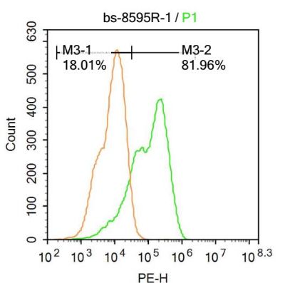
U-937 cells were fixed with 4% PFA for 10min at room temperature,permeabilized with 90% ice-cold methanol for 20 min at room temperature,and incubated in 5% BSA blocking buffer for 30 min at room temperature. Cells were then stained with C9orf72 Antibody(SL8595R) at 1:100 dilution in blocking buffer and incubated for 30 min at room temperature, washed twice with 2%BSA in PBS, followed by secondary antibody incubation for 40 min at room temperature. Acquisitions of 20,000 events were performed.Cells stained with primary antibody (green), and isotype control (orange).
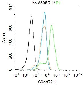
Blank control:Hela.
Primary Antibody (green line): Rabbit Anti-C9orf72 antibody (SL8595R)
Dilution: 1μg /10^6 cells;
Isotype Control Antibody (orange line): Rabbit IgG .
Secondary Antibody : Goat anti-rabbit IgG-FITC
Dilution: 1μg /test.
Protocol
The cells were fixed with 4% PFA (10min at room temperature)and then permeabilized with 0.1% PBST for 20 min at room temperature. The cells were then incubated in 5%BSA to block non-specific protein-protein interactions for 30 min at room temperature .Cells stained with Primary Antibody for 30 min at room temperature. The secondary antibody used for 40 min at room temperature. Acquisition of 20,000 events was performed.
|
|
|