Home > Product > Antibody > Rabbit Anti-HIPPI antibody
Dermal papilla derived protein 8; Dermal papilla-derived protein 8; DERP 8; DERP8; Esrrbl 1; Esrrbl1; ESRRBL1 protein; Estrogen related receptor beta like 1; Estrogen related receptor beta like protein 1; Estrogen-related receptor beta like 1; Estrogen-re
Cat:
SL11697R
Species Reactivity:
Human,Mouse,Rat,(predicted: Dog,Cow,Horse,Rabbit,)
Immunogen:
KLH conjugated synthetic peptide derived from human HIPPI:331-429/429
Format:
Liquid
Storage instructions:
Shipped at 4℃. Store at -20 °C for one year. Avoid repeated freeze/thaw cycles.
Concentration:
1mg/ml
Clonality:
Polyclonal
Isotype:
IgG
Applications:
WB=1:500-2000ELISA=1:5000-10000IHC-P=1:100-500IHC-F=1:100-500ICC=1:100-500IF=1:100-500(Paraffin sections need to do antigen repair)not yet tested in other applications.optimal dilutions/concentrations should be determined by the end user.
Host:
Rabbit
Product Overview:
Sample: Lymph node (Mouse) Lysate at 40 ugLung (Mouse) Lysate at 40 ugPrimary: Anti- HIPPI (SL11697R) at 1/1000 dilutionSecondary: IRDye800CW Goat Anti-Rabbit IgG at 1/20000 dilutionPredicted band size: 49 kDObserved band size: 52 kDSample: Thymus (Mouse) Lysate at 40 ugLiver (Mouse) Lysate at 40 ugPrimary: Anti- HIPPI (SL11697R) at 1/1000 dilutionSecondary: IRDye800CW Goat Anti-Rabbit IgG at 1/20000 dilutionPredicted band size: 49 kDObserved band size: 53 kDSample: A431(Human) Cell Lysate at 30 ugPrimary: Anti- HIPPI (SL11697R) at 1/1000 dilutionSecondary: IRDye800CW Goat Anti-Rabbit IgG at 1/20000 dilutionPredicted band size: 49 kDObserved band size: 52 kDParaformaldehyde-fixed, paraffin embedded (rat brain); Antigen retrieval by boiling in sodium citrate buffer (pH6.0) for 15min; Block endogenous peroxidase by 3% hydrogen peroxide for 20 minutes; Blocking buffer (normal goat serum) at 37°C for 30min; Antibody incubation with (HIPPI) Polyclonal Antibody, Unconjugated (SL11697R) at 1:200 overnight at 4°C, followed by operating according to SP Kit(Rabbit) (sp-0023) instructionsand DAB staining.
More
Unit:
Price: $
Product PDFs
Datasheet:


Programmed neuronal cell death is a feature of neurodegenerative disorders such as Alzheimer's and Huntington's disease, which occur later in human life. Huntington’s disease at the molecular and cell level is characterized by polyglutamine expansion of the protein huntingtin (Htt) that leads to apoptotis-mediated neurodegenerative loss of medium spiny neurons throughout the striatum. Polyglutamine expansion reduces the level of association between Hip-1 and Htt, thereby increasing levels of free Hip-1 that then can be the candidate protein Hippi (Hip-1 protein interactor). The Hippi-Hip-1 heterodimer is a pro-apoptotic complex that recruits procaspase-8 and initiates caspase-8 activation, which may contribute to the neuronal cell death observed in individuals diagnosed with Huntington’s disease. The human hippi gene maps to chromosome 3q13.13 and encodes a 429 amino acid protein.

Function:
Required for the formation of cilia. Plays an indirect role in sonic hedgehog signaling, cilia being required for all activity of the hedgehog pathway (By similarity). Has pro-apoptotic function via its interaction with HIP1, leading to recruit caspase-8 (CASP8) and trigger apoptosis. Has the ability to bind DNA sequence motif 5'-AAAGACATG-3' present in the promoter of caspase genes such as CASP1, CASP8 and CASP10, suggesting that it may act as a transcription regulator; however the relevance of such function remains unclear.

Subunit:
Component of IFT complex B composed of IFT88, IFT57, TRAF3IP1, IFT52, IFT27, HSPB11 and IFT20. Interacts with IFT20 and IFT88. Interacts with BLOC1S2 and RYBP. Interacts with HOMER1, possibly leading to prevent pro-apoptotic effects of IFT57 (By similarity). Interacts with HIP1. In normal conditions, it poorly interacts with HIP1, HIP1 being strongly associated with HTT. However, in mutant HTT proteins with a long poly-Gln region, interaction between HTT and HIP1 is inhibited, promoting the interaction between HIP1 and IFT57, leading to apoptosis. Interacts with SFAR. Interacts with chicken anemia virus protein apoptin.

Subcellular Location:
Cytoplasm, cytoskeleton, cilium basal body (By similarity). Note=Concentrates within the inner segment of cilia

Tissue Specificity:
Present in many tissues such as brain, thymus, lymph node, lung, liver, skin and kidney (at protein level).

Similarity:
Belongs to the IFT57 family.

SWISS:
Q9NWB7

Gene ID:
55081

Database links:

Entrez Gene: 55081 Human

Entrez Gene: 73916 Mouse

Omim: 606621 Human

SwissProt: Q9NWB7 Human

SwissProt: Q8BXG3 Mouse

Unigene: 412196 Human

Unigene: 241276 Mouse

Unigene: 404371 Mouse


Picture

Sample:
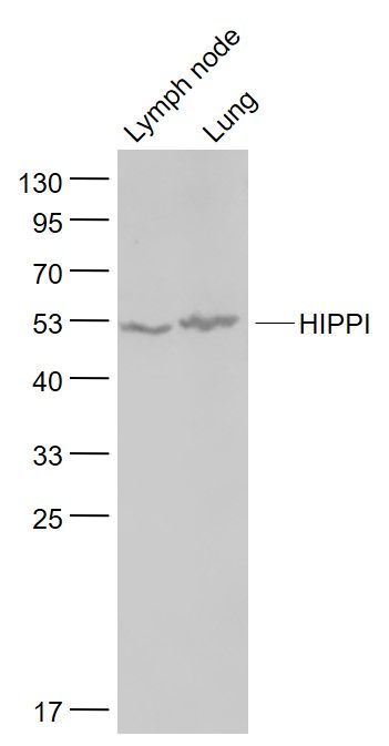
Lymph node (Mouse) Lysate at 40 ug
Lung (Mouse) Lysate at 40 ug
Primary: Anti- HIPPI (SL11697R) at 1/1000 dilution
Secondary: IRDye800CW Goat Anti-Rabbit IgG at 1/20000 dilution
Predicted band size: 49 kD
Observed band size: 52 kD
Sample:
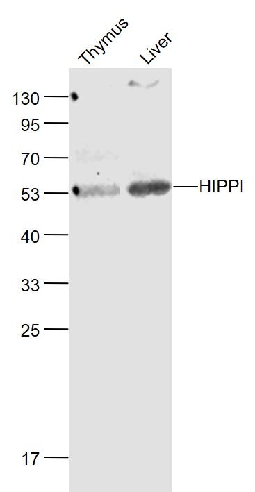
Thymus (Mouse) Lysate at 40 ug
Liver (Mouse) Lysate at 40 ug
Primary: Anti- HIPPI (SL11697R) at 1/1000 dilution
Secondary: IRDye800CW Goat Anti-Rabbit IgG at 1/20000 dilution
Predicted band size: 49 kD
Observed band size: 53 kD
Sample:
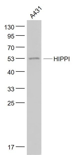
A431(Human) Cell Lysate at 30 ug
Primary: Anti- HIPPI (SL11697R) at 1/1000 dilution
Secondary: IRDye800CW Goat Anti-Rabbit IgG at 1/20000 dilution
Predicted band size: 49 kD
Observed band size: 52 kD
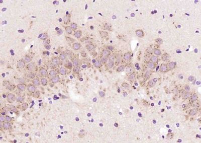
Paraformaldehyde-fixed, paraffin embedded (rat brain); Antigen retrieval by boiling in sodium citrate buffer (pH6.0) for 15min; Block endogenous peroxidase by 3% hydrogen peroxide for 20 minutes; Blocking buffer (normal goat serum) at 37°C for 30min; Antibody incubation with (HIPPI) Polyclonal Antibody, Unconjugated (SL11697R) at 1:200 overnight at 4°C, followed by operating according to SP Kit(Rabbit) (sp-0023) instructionsand DAB staining.
Product Feedback Wall
Message :
Your Email :
Copyright © 2007-2018 Sunlong Medical All Rights Reserved.