Specific References (4) | SL10159R has been referenced in 4 publications.
[IF=6.832] Aussel, Clotilde. et al. IL-1β primed mesenchymal stromal cells moderate hemorrhagic shock-induced organ injuries. Stem Cell Res Ther. 2021 Dec;12(1):1-16 FC ; Rat.
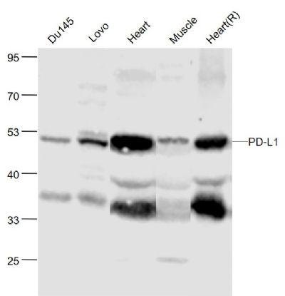
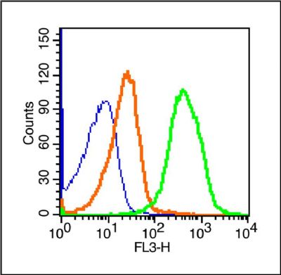
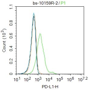
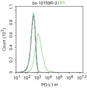
[IF=3.098] Junli Zhang. et al. KCNQ1OT1 contributes to sorafenib resistance and programmed death‑ligand‑1‑mediated immune escape via sponging miR‑506 in hepatocellular carcinoma cells. Int J Mol Med. 2020 Nov;46(5):1794-364 WB,IHC,FC ; Human.
[IF=4.086] Yi-Ru Pan. et al. Comprehensive Evaluation of Immune-Checkpoint DNA Cancer Vaccines in a Rat Cholangiocarcinoma Model. Vaccines-Basel. 2020 Dec;8(4):703 IHC ; Rat.
[IF=4.932] Shuling Zhang. et al. A novel mechanism of lung cancer inhibition by methionine enkephalin through remodeling the immune status of the tumor microenvironment. Int Immunopharmacol. 2021 Oct;99:107999 IF ; Mouse.