Sample:
GFP-Tagged Fusion Protein Overexpression E.coli Lysate (Cat#: SL33009P) at 2 ug
Primary: Anti-GFP-Tag (SLM51005M) at 1/2000 ~ 1/10000 dilution
Secondary: IRDye800CW Goat Anti-Mouse IgG at 1/20000 dilution
Predicted band size: 28 kD
Observed band size: 28 kD
Sample:
GFP-Tagged Fusion Protein Overexpression E.coli Lysate (Cat#: SL33009P) at 4 ug
Primary: Anti-GFP-Tag (SLM51005M) at 1/2000 ~ 1/10000 dilution
Secondary: IRDye800CW Goat Anti-Mouse IgG at 1/20000 dilution
Predicted band size: 28 kD
Observed band size: 28 kD
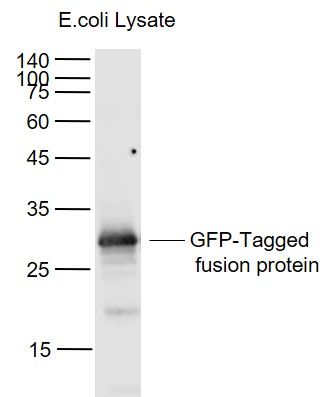
Sample:
Lane 1: GFP-Tagged fusion protein (full length) Overexpression E.coli Lysate (Cat#: SL33009P) at 4 ug
Primary: Anti-GFP-Tag (SLM51005M) at 1/1000 dilution
Secondary: IRDye800CW Goat Anti-Mouse IgG at 1/20000 dilution
Predicted band size: 28 kD
Observed band size: 28 kD
|