Home > Product > Antibody > Rabbit Anti-P14ARF/FITC Conjugated antibody
CDKN2A; p16INK4a; INK4; P16INK4; ARF; CCM2; CDK4 Inhibitor; CDK4 inhibitor p16 INK4; CDK4I; CDKN2; CDKN2A; Cell cycle negative regulator beta; CMM2; Cyclin dependent kinase 4 inhibitor A; Cyclin dependent kinase inhibitor 2A (melanoma p16 inhibits CDK4);
Cat:
SL4592R-FITC
Species Reactivity:
Human,
Immunogen:
KLH conjugated synthetic peptide derived from human P16/CDKN2A
Format:
Lyophilized or Liquid
Storage instructions:
Store at -20 °C for one year. Avoid repeated freeze/thaw cycles. The lyophilized antibody is stable at room temperature for at least one month and for greater than a year when kept at -20°C. When reconstituted in sterile pH 7.4 0.01M PBS or diluent of ant
Buffer:
0.01M TBS(pH7.4) with 1% BSA, 0.03% Proclin300 and 50% Glycerol.
Concentration:
1mg/ml
Clonality:
Polyclonal
Isotype:
IgG
Applications:
Flow-Cyt=1ug/TestIF=1:50-200not yet tested in other applications.optimal dilutions/concentrations should be determined by the end user.
Host:
Rabbit
Calculated MW:
15kDa
Product Overview:
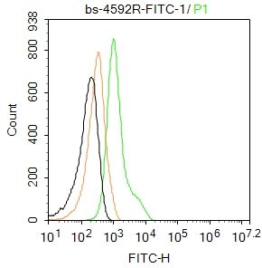
Blank control: Hela. Primary Antibody (green line): Rabbit Anti-P14ARF antibody (SL4592R-FITC) Dilution: 1μg /10^6 cells; Isotype Control Antibody (orange line): Rabbit IgG . ProtocolThe cells were fixed with 4% PFA (10min at room temperature)and then permeabilized with 90% ice-cold methanol for 20 min at -20℃. The cells were then incubated in 5%BSA to block non-specific protein-protein interactions for 30 min at at room temperature .Cells stained with Primary Antibody for 30 min at room temperature.Acquisition of 20,000 events was performed.
More
Unit:
Price: $
Product PDFs
Datasheet:


background:
The CDKN2A locus gives rise to 2 distinct transcripts from different promoters. The transcripts have been designated p16(INK4A) and p14(ARF). This chromosomal region undergoes a number of inversions, translocations, heterozygous deletions, and homozygous deletions in a variety of malignant cell lines including those from glioma, non-small cell lung cancer, leukemia, and melanoma. Deletion of the region containing CDKN2A is found in more than half of all melanoma cell lines. In spite of the structural and some functional differences, all the proteins encoded by the CDKN2A gene are involved in cell cycle G1 control.

Function:
Capable of inducing cell cycle arrest in G1 and G2 phases. Acts as a tumor suppressor. Binds to MDM2 and blocks its nucleocytoplasmic shuttling by sequestering it in the nucleolus. This inhibits the oncogenic action of MDM2 by blocking MDM2-induced degradation of p53 and enhancing p53-dependent transactivation and apoptosis. Also induces G2 arrest and apoptosis in a p53-independent manner by preventing the activation of cyclin B1/CDC2 complexes. Binds to BCL6 and down-regulates BCL6-induced transcriptional repression. Binds to E2F1 and MYC and blocks their transcriptional activator activity but has no effect on MYC transcriptional repression. Binds to TOP1/TOPOI and stimulates its activity. This complex binds to rRNA gene promoters and may play a role in rRNA transcription and/or maturation. Interacts with NPM1/B23 and promotes its polyubiquitination and degradation, thus inhibiting rRNA processing. Interacts with COMMD1 and promotes its 'Lys63'-linked polyubiquitination. Interacts with UBE2I/UBC9 and enhances sumoylation of a number of its binding partners including MDM2 and E2F1. Binds to HUWE1 and represses its ubiquitin ligase activity. May play a role in controlling cell proliferation and apoptosis during mammary gland development.

Subunit:
Does not interact with cyclins, CDK1, CDK2, CDK4, CDK5 or CDK6. Binds to BCL6, E2F1, HUWE1, MDM2, MYC, NPM1/B23, TOP1/TOPOI and UBE2I/UBC9. Interacts with TBRG1 and COMMD1. Interacts with CDKN2AIP and E4F1.

Subcellular Location:
Nucleus, nucleolus. Nucleus, nucleoplasm.

Post-translational modifications:
Ubiquitinated in normal cells by TRIP12 via the ubiquitin fusion degradation (UFD) pathway, a process that mediates ubiquitination at the N-terminus, regardeless of the absence of lysine residues. Ubiquitination leads to its degradation. In cancer cells, however, TRIP12 is located in a different cell compartment, preventing ubiquitination and degradation.

Database links:

Entrez Gene: 1029 Human

Omim: 600160 Human

SwissProt: P42771 Human

SwissProt: Q8N726 Human

Unigene: 512599 Human


Important Note:
This product as supplied is intended for research use only, not for use in human, therapeutic or diagnostic applications.

细胞周期失控是癌变的重要原因。p16是近年来发现的第一个直接参与细胞周期调控的抑癌基因,其表达产物为p16蛋白.
p16基因是一种重要的抑癌基因,在正常细胞中起负反馈作用,当p16基因突变或丢失时,细胞增殖失去控制使细胞无限制地增殖。
p16主要功能是通过抑制CDK4而阻止细胞由G1期进入S期,使细胞增殖受到限制。用于各种恶性肿瘤如肺癌、恶黑、乳腺癌的研究。目前的研究细胞周期依赖激酶抑制p16INK4a蛋白在宫颈上皮内病变(CIN)中作为一个新标记物.
p16INK4a的过表达与HPV E7区(病毒早期蛋白即病毒致癌基因编码区)活性有密切相关性。
p16/CDKN2基因是新近发现的肿瘤抑制基因,已有研究表明该基因在许多肿瘤出现缺失、突变或重排现象. Picture

Blank control: Hela.
Primary Antibody (green line): Rabbit Anti-P14ARF antibody (SL4592R-FITC)
Dilution: 1μg /10^6 cells;
Isotype Control Antibody (orange line): Rabbit IgG .
Protocol
The cells were fixed with 4% PFA (10min at room temperature)and then permeabilized with 90% ice-cold methanol for 20 min at -20℃. The cells were then incubated in 5%BSA to block non-specific protein-protein interactions for 30 min at at room temperature .Cells stained with Primary Antibody for 30 min at room temperature.Acquisition of 20,000 events was performed.
Product Feedback Wall
Message :
Your Email :
Copyright © 2007-2018 Sunlong Medical All Rights Reserved.