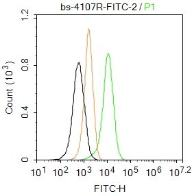
Blank control: Raji.
Primary Antibody (green line): Rabbit Anti-MHC antibody (SL4107R-FITC)
Dilution: 2μg /10^6 cells;
Isotype Control Antibody (orange line): Rabbit IgG .
Protocol
The cells were fixed with 4% PFA (10min at room temperature)and then permeabilized with PBST for 20 min at room temperature. The cells were then incubated in 5%BSA to block non-specific protein-protein interactions for 30 min at at room temperature .Cells stained with Primary Antibody for 30 min at room temperature. Acquisition of 20,000 events was performed.
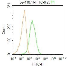
Blank control: Mouse spleen.
Primary Antibody (green line): Rabbit Anti-MHC Class II/HLA DMB/FITC Conjugated antibody (SL4107R-FITC)
Dilution: 0.2μg /10^6 cells;
Isotype Control Antibody (orange line): Rabbit IgG-FITC .
Protocol
The cells were fixed with 4% PFA (10min at room temperature)and then permeabilized with 0.1% PBST for 20 min at-20℃.The cells were then incubated in 5% BSA to block non-specific protein-protein interactions for 30 min at room temperature. The cells were stained with Primary Antibody for 30 min at room temperature. Acquisition of 20,000 events was performed.