background:
The protein encoded by this gene is a cell-surface glycoprotein involved in cell-cell interactions, cell adhesion and migration. It is a receptor for hyaluronic acid (HA) and can also interact with other ligands, such as osteopontin, collagens, and matrix metalloproteinases (MMPs). This protein participates in a wide variety of cellular functions including lymphocyte activation, recirculation and homing, hematopoiesis, and tumor metastasis. Transcripts for this gene undergo complex alternative splicing that results in many functionally distinct isoforms, however, the full length nature of some of these variants has not been determined. Alternative splicing is the basis for the structural and functional diversity of this protein, and may be related to tumor metastasis. [provided by RefSeq, Jul 2008].
Function:
Receptor for hyaluronic acid (HA). Mediates cell-cell and cell-matrix interactions through its affinity for HA, and possibly also through its affinity for other ligands such as osteopontin, collagens, and matrix metalloproteinases (MMPs). Adhesion with HA plays an important role in cell migration, tumor growth and progression. Also involved in lymphocyte activation, recirculation and homing, and in hematopoiesis. Altered expression or dysfunction causes numerous pathogenic phenotypes. Great protein heterogeneity due to numerous alternative splicing and post-translational modification events.
Subunit:
Interacts with PKN2 (By similarity). Interacts with HA, as well as other glycosaminoglycans, collagen, laminin, and fibronectin via its N-terminal segment. Interacts with ANK, the ERM proteins (VIL2, RDX and MSN), and NF2 via its SLCterminal segment.
Subcellular Location:
Membrane; Single-pass type I membrane protein. Note=Colocalizes with actin in membrane protrusions at wounding edges.
Tissue Specificity:
Isoform 10 (epithelial isoform) is expressed by cells of epithelium and highly expressed by carcinomas. Expression is repressed in neuroblastoma cells.
Post-translational modifications:
Proteolytically cleaved in the extracellular matrix by specific proteinases (possibly MMPs) in several cell lines and tumors.
N- and O-glycosylated. O-glycosylation contains more-or-less-sulfated chondroitin sulfate glycans, whose number may affect the accessibility of specific proteinases to their cleavage site(s). It is uncertain if O-glycosylation occurs on Thr-637 or Thr-638.
Phosphorylated; activation of PKC results in the dephosphorylation of Ser-706 (constitutive phosphorylation site), and the phosphorylation of Ser-672.
Similarity:
Contains 1 Link domain.
Database links:
Entrez Gene: 281057 Cow
Entrez Gene: 960 Human
Entrez Gene: 12505 Mouse
Entrez Gene: 100301546 Rabbit
Entrez Gene: 25406 Rat
Omim: 107269 Human
SwissProt: Q29423 Cow
SwissProt: P16070 Human
SwissProt: P15379 Mouse
SwissProt: P26051 Rat
Unigene: 502328 Human
Unigene: 423621 Mouse
Unigene: 1120 Rat
Important Note:
This product as supplied is intended for research use only, not for use in human, therapeutic or diagnostic applications.
CD44是一个重要的细胞表面粘附分子。它在许多类型的人类细胞上都表达,在很多生理和病理过程中有复杂的作用,例如细胞迁移和在肿瘤细胞的生长调控中。CD44传递着诸多的细胞通路,有研究者认为CD44是一个新的信号通路蛋白。CD44介导淋巴细胞与血管内皮细胞包括粘膜组织中高内皮小静脉的粘连,这可能是通过激活其它粘附分子而间接造成的。CD44也是肿瘤细胞浸润的重要因素。
| Picture |
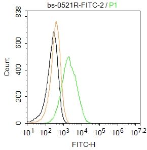
Blank control:RAW264.7.
Primary Antibody (green line): Rabbit Anti-CD44 antibody (SL0521R-FITC)
Dilution: 2μg /10^6 cells;
Isotype Control Antibody (orange line): Rabbit IgG .
Protocol
The cells were incubated in 5%BSA to block non-specific protein-protein interactions for 30 min at room temperature .Cells stained with Primary Antibody for 30 min at room temperature. Acquisition of 20,000 events was performed.
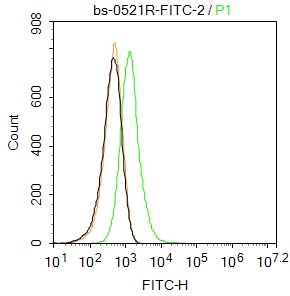
Blank control:Hela.
Primary Antibody (green line): Rabbit Anti-CD44 antibody (SL0521R-FITC)
Dilution: 2μg /10^6 cells;
Isotype Control Antibody (orange line): Rabbit IgG .
Protocol
The cells were incubated in 5%BSA to block non-specific protein-protein interactions for 30 min at room temperature .Cells stained with Primary Antibody for 30 min at room temperature. Acquisition of 20,000 events was performed.
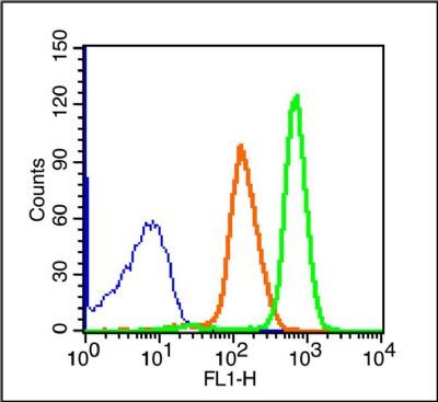
Blank control (blue line): Rabbit spleen cells(blue).
Primary Antibody (green line): Rabbit Anti-CD44/FITC Conjugated antibody (SL0521R-FITC)
Dilution: 1μg /10^6 cells;
Isotype Control Antibody (orange line): Rabbit IgG-FITC.
Protocol
The cells were fixed with 70% ice-cold methanol overnight at 4℃ . The cells were then incubated in 1 X PBS/2%BSA/10% goat serum to block non-specific protein-protein interactions followed by the antibody for 15 min at room temperature. Cells stained with Primary Antibody for 30 min at room temperature.Acquisition of 20,000 events was performed.
|
|
|