[IF=3.269] Lin Wang. et al. Suppression of NOD-like receptor protein 3 inflammasome activation and macrophage M1 polarization by hederagenin contributes to attenuation of sepsis-induced acute lung injury in rats. Bioengineered. 2022;13(3):7262-7276 WB ; Rat,Human.
[IF=2.1] Hu, Hanhua, et al. "Drug-induced apoptosis of Echinococcus granulosus protoscoleces." Parasitology research 109.2 (2011): 453-459. IHSLCP ; Others.
[IF=4.411] Kathleen Pappritz. et al. Colchicine prevents disease progression in viral myocarditis via modulating the NLRP3 inflammasome in the cardiosplenic axis. 2022 Feb 17 FC ; Mouse.
[IF=5.279] Lei Wang. et al. Lactobacillus plantarum DP189 Reduces α-SYN Aggravation in MPTP-Induced Parkinson’s Disease Mice via Regulating Oxidative Damage, Inflammation, and Gut Microbiota Disorder. J Agr Food Chem. 2022;70(4):1163–1173 WB ; Mouse.
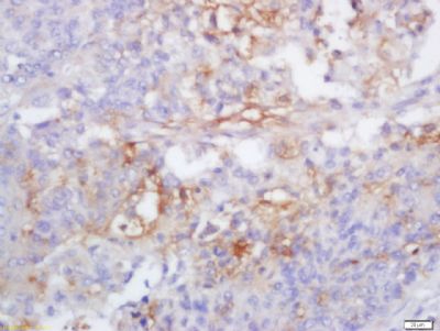
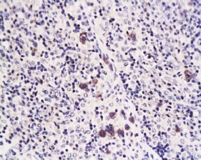
[IF=3.337] Ziyu Meng. et al. Sesamin promotes apoptosis and pyroptosis via autophagy to enhance antitumour effects on murine T-cell lymphoma. J Pharmacol Sci. 2021 Nov;147:260 WB,IHC ; Mouse.
[IF=2.615] Wei Yu. et al. Curcumin suppresses doxorubicin-induced cardiomyocyte pyroptosis via a PI3K/Akt/mTOR-dependent manner. Cardiovasc Diagn The. 2020 Aug; 10(4): 752–769 WB ; Mouse.
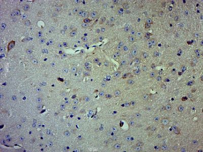
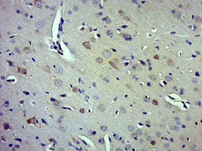
[IF=4.362] Zhou Wen. et al. Enhanced Autolysosomal Function Ameliorates the Inflammatory Response Mediated by the NLRP3 Inflammasome in Alzheimer’s Disease. Front Aging Neurosci. 2021 Feb;13:46 WB ; Mouse.
[IF=3.266] Guan X et al. Estrogen deficiency aggravates apical periodontitis by regulating NLRP3/caspase-1/IL-1β axis. Am J Transl Res. 2020 Feb 15;12(2):660-671. WB,IHC ; human.
[IF=2.842] Guangrui Chai. et al. NLRP3 Blockade Suppresses Pro-Inflammatory and Pro-Angiogenic Cytokine Secretion in Diabetic Retinopathy. Diabet Metab Synd Ob. 2020; 13: 3047–3058 WB ; Human.
[IF=1.813] Zhu Hui-Zheng. et al. Xiaoyaosan Exerts Therapeutic Effects on the Colon of Chronic Restraint Stress Model Rats via the Regulation of Immunoinflammatory Activation Induced by the TLR4/NLRP3 Inflammasome Signaling Pathway. Evid-Based Compl Alt. 2021;2021:6673538 WB ; Rat.
[IF=4.321] Shu-Min Yeet al. Silencing of Gasdermin D by siRNA-Loaded PEI-Chol Lipopolymers Potently Relieves Acute Gouty Arthritis through Inhibiting Pyroptosis. Mol Pharm
. 2020 Jul 6. WB ; mouse.
[IF=3.743] Chen Y et al. Curcumin attenuates potassium oxonate-induced hyperuricemia and kidney inflammation in mice. Biomed Pharmacother. 2019 Jul 27;118:109195. WB ; Mouse.
[IF=3.974] Liao J et al. Inhibition of Caspase-1-dependent pyroptosis attenuates copper-induced apoptosis in chicken hepatocytes.Ecotoxicol Environ Saf. 2019 Jun 15;174:110-119. WB ; Chicken.
[IF=4.263] Wu X et al.The role of Ca 2+ in acid-sensing ion channel 1a-mediated chondrocyte pyroptosis in rat adjuvant arthritis.(2018) Lab Invest. WB ; Rat.
[IF=3.23] Daverey, Amita, and Sandeep K. Agrawal. "Curcumin alleviates oxidative stress and mitochondrial dysfunction in astrocytes." Neuroscience 333 (2016): 92-103. WB ; Human.
[IF=3.32] Yao, Xiao, et al. "Estrogen-provided cardiac protection following burn trauma is mediated through a reduction in mitochondria-derived DAMPs." Am J Physiol Heart Circ Physiol 306.6 (2014): H882-H894. IHSLCP ;
[IF=4.6] Wang, Shaolan, et al. "Xenobiotic receptor PXR regulates innate immunity via activation of NLRP3 inflammasome in vascular endothelial cells." Journal of Biological Chemistry (2014): jbc-M114. WB ; Human.