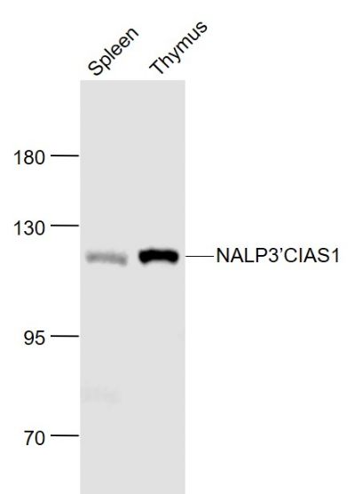
[IF=8.469] Pang, Qi. et al. Irisin protects against vascular calcification by activating autophagy and inhibiting NLRP3-mediated vascular smooth muscle cell pyroptosis in chronic kidney disease. Cell Death Dis. 2022 Mar;13(3):1-15 IF ; Mouse.
[IF=3.04] Niu X et al. Harmine mitigates LPS-induced acute kidney injury through inhibition of the TLR4-NF-κB/NLRP3 inflammasome signalling pathway in mice. Eur J Pharmacol. 2019 Apr 15;849:160-169. IHSLCP&WB ; Mouse.
[IF=4.225] Liu P et al. Harmine Ameliorates Cognitive Impairment by Inhibiting NLRP3 Inflammasome Activation and Enhancing the BDNF/TrkB Signaling Pathway in STZ-Induced Diabetic Rats. Front Pharmacol. 2020 May 1;11:535. WB/IHC/IF ; Rat.
[IF=2.615] Wei Yu. et al. Curcumin suppresses doxorubicin-induced cardiomyocyte pyroptosis via a PI3K/Akt/mTOR-dependent manner. Cardiovasc Diagn The. 2020 Aug; 10(4): 752–769 WB ; Mouse.
[IF=8.469] Zheng, Jie. et al. A novel function of NLRP3 independent of inflammasome as a key transcription factor of IL-33 in epithelial cells of atopic dermatitis. Cell Death Dis. 2021 Sep;12(10):1-12 IHC ; mouse.
[IF=3.683] Shi, Chenxi. et al. Dihydromyricetin alleviates Escherichia coli lipopolysaccharide-induced hepatic injury in chickens by inhibiting the NLRP3 inflammasome. Vet Res. 2022 Dec;53(1):1-16 WB ; Chicken.
[IF=3.59] Jie Ma. et al. The Effect of MaxiK Channel on Regulating the Activation of NLRP3 Inflammasome in Rats of Blast-induced Traumatic Brain Injury. Neuroscience. 2022 Feb;482:132 IHC ; Rat.
[IF=4.268] Xiping Yang. et al. The piperine derivative HJ105 inhibits Aβ1-42-induced neuroinflammation and oxidative damage via the Keap1-Nrf2-TXNIP axis. Phytomedicine. 2021 Apr;:153571 WB ; Rat.
[IF=3.056] Carmen Hummel. et al. Expression and Cell Type-specific Localization of Inflammasome Sensors in the Spinal Cord of SOD1(G93A) Mice and Sporadic Amyotrophic lateral sclerosis Patients. Neuroscience. 2021 Mar;: IHC ; Mouse.
[IF=4.966] Hui Che. et al. Melatonin exerts neuroprotective effects by inhibiting neuronal pyroptosis and autophagy in STZ‐induced diabetic mice. Faseb J. 2020 Oct;34(10):14042-14054 WB,IHC ; Mouse, Human.
[IF=3.9] Dong Zhixia. et al. Adiponectin Inhibits NLRP3 Inflammasome Activation in Nonalcoholic Steatohepatitis via AMPK-JNK/ErK1/2-NFκB/ROS Signaling Pathways. Front Med-Lausanne. 2020 Nov;7:705 WB ; Mouse.
[IF=4.362] Zhou Wen. et al. Enhanced Autolysosomal Function Ameliorates the Inflammatory Response Mediated by the NLRP3 Inflammasome in Alzheimer’s Disease. Front Aging Neurosci. 2021 Feb;13:46 WB ; Mouse.
[IF=3.647] Guan L et al. Puerarin ameliorates retinal ganglion cell damage induced by retinal ischemia/reperfusion through inhibiting the activation of TLR4/NLRP3 inflammasome. Life Sci
. 2020 Sep 1;256:117935. WB ; Rat.
[IF=6.304] Hanwen Li et al. P2Y 14 receptor has a critical role in acute gouty arthritis by regulating pyroptosis of macrophages. Cell Death Dis.2020 May 26;11(5):394. WB ; Rat.
[IF=3.361] Yang X et al. HJ22, a Novel derivative of piperine, Attenuates ibotenic acid-induced cognitive impairment, oxidativestress, apoptosis and inflammation via inhibiting the protein-protein interaction of Keap1-Nrf2. Int Immunopharmacol. 2020 Mar 16;83:106383. IHC,WB ; rat.
[IF=3.266] Guan X et al. Estrogen deficiency aggravates apical periodontitis by regulating NLRP3/caspase-1/IL-1β axis. Am J Transl Res. 2020 Feb 15;12(2):660-671. WB,IHC ; human.
[IF=1.773] Qu XY et al. XingNaoJing injections protect against cerebral ischemia/reperfusion injury and alleviate blood-brain barrier disruption in rats, through an underlying mechanism of NLRP3 inflammasomes suppression. Chin J Nat Med. 2019 Jul;17(7):498-505. WB&IHSLCP ; Rat.
[IF=3.118] Peng XP et al. The protective effect of oleanolic acid on NMDA-induced MLE-12 cells apoptosis and lung injury in mice by activating SIRT1 and reducing NF-κB acetylation.Int Immunopharmacol. 2019 May;70:520-529. WB ; Mouse.
[IF=5.589] WB + ELISA ; Mouse.
[IF=1.719] Qu XY et al. Astragaloside IV protects against cisplatin-induced liver and kidney injury via autophagy-mediated inhibition of NLRP3 in rats.The Journal of Toxicological Sciences,2019 44(3), 167–175. IHSLCP&WB ; Rat.
[IF=4.263] Wu X et al.The role of Ca 2+ in acid-sensing ion channel 1a-mediated chondrocyte pyroptosis in rat adjuvant arthritis.(2018) Lab Invest. WB ; Rat.
[IF=6.187] Debye et al. Neurodegeneration and NLRP3 inflammasome expression in the anterior thalamus of SOD1(G93A) ALS mice. (2016) Brain.Pathol. IHSLCP,IHSLCP-IF ; Mouse.
[IF=5.168] Yu, Chengyuan, et al. "Chronic obstructive sleep apnea promotes aortic remodeling in canines through miR-145/Smad3 signaling pathway." Oncotarget 8.23 (2017): 37705. WB ; Dog.
[IF=5.4] Zendedel, Adib, et al. "Estrogen Attenuates Local Inflammasome Expression and Activation after Spinal Cord Injury." Molecular Neurobiology (2017): 1-12. WB, IF(IHSLCP) ; Rat.
[IF=4.21] Zhou, Tianwei E., et al. "Choroidal Involution Is Associated with a Progressive Degeneration of the Outer Retinal Function in a Model of Retinopathy of Prematurity: Early Role for IL-1β." The American Journal of Pathology (2016). IHSLCF ; Rat.
[IF=5.29] Zendedel, Adib, et al. "Activation and Regulation of NLRP3 Inflammasome by Intrathecal Application of SDF-1a in a Spinal Cord Injury Model." Molecular Neurobiology (2015): 1-13. WB ; Rat.
[IF=4.095] Slowik A et al. Impact of steroid hormones E2 and P on the NLRP3/ASC/Casp1 axis in primary mouse astroglia and BSLV2 cells after in vitro hypoxia.J Steroid Biochem Mol Biol. 2018 Oct;183:18-26. WB&ICF ; Murine.
[IF=1.813] Zhu Hui-Zheng. et al. Xiaoyaosan Exerts Therapeutic Effects on the Colon of Chronic Restraint Stress Model Rats via the Regulation of Immunoinflammatory Activation Induced by the TLR4/NLRP3 Inflammasome Signaling Pathway. Evid-Based Compl Alt. 2021;2021:6673538 WB ; Rat.
[IF=2.447] Zijie Wei. et al. KDM2B overexpression prevents myocardial ischemia‑reperfusion injury in rats through regulating inflammatory response via the TLR4/NF‑κB p65 axis. Exp Ther Med. 2022 Feb;23(2):1-8 WB ; Rat.
[IF=5.396] Shi-Yong Zhu. et al. Lycopene Ameliorates Atrazine-Induced Pyroptosis in Spleen via Suppressing the Ox-mtDNA/Nlrp3 Inflammasome Pathway. Food Funct. 2021 Nov;: IF ; Mouse.
[IF=3.921] Meiyun Cai. et al. Kaemperfol alleviates pyroptosis and microglia-mediated neuroinflammation in Parkinson's disease via inhibiting p38MAPK/NF-κB signaling pathway. Neurochem Int. 2021 Nov;:105221 WB ; Rat.
[IF=6.6] Maryam Baazm. et al. Regulation of Inflammasomes by Application of Omega-3 Polyunsaturated Fatty Acids in a Spinal Cord Injury Model. Cells-Basel. 2021 Nov;10(11):3147 WB ; Rat.
[IF=6.543] Wen Lianghe. et al. Melatonin Exerts Cardioprotective Effects by Inhibiting NLRP3 Inflammasome-Induced Pyroptosis in Mice following Myocardial Infarction. Oxid Med Cell Longev. 2021;2021:5387799 WB,IF ; mouse.
[IF=7.561] You Zhou. et al. Overexpression of lncRNA TUG1 Alleviates NLRP3 Inflammasome-Mediated Cardiomyocyte Pyroptosis Through Targeting the miR-186-5p/XIAP Axis in Coronary Microembolization-Induced Myocardial Damage. Front Immunol. 2021; 12: 637598 WB ; Rat.
[IF=6.6] Chinmaya Panda. et al. Aggregated Tau-PHF6 (VQIVYK) Potentiates NLRP3 Inflammasome Expression and Autophagy in Human Microglial Cells. Cells-Basel. 2021 Jul;10(7):1652 ICC ; Human.
[IF=2.678] Huan Xiao. et al. Effects of Mycobacterium vaccae Aerosol Inhalation on Airway Inflammation in Asthma Mouse Model. 2021 May 04 IHC ; Mouse.
[IF=1.695] Yaru Huang. et al. Dexmedetomidine attenuates myocardial ischemia-reperfusion injury in vitro by inhibiting NLRP3 Inflammasome activation. Bmc Anesthesiol. 2021 Dec;21(1):1-12 WB ; Rat.
[IF=3.758] Lei Xianghong. et al. Micheliolide Attenuates Lipopolysaccharide-Induced Inflammation by Modulating the mROS/NF-κB/NLRP3 Axis in Renal Tubular Epithelial Cells. Mediat Inflamm. 2020;2020:3934769 WB ; Rat.
[IF=3.701] Ying Sun. et al. Neuroprotective effects of natural cordycepin on LPS-induced Parkinson’s disease through suppressing TLR4/NF-κB/NLRP3-mediated pyroptosis. J Funct Foods. 2020 Dec;75:104274 WB ; Mouse.
[IF=7.919] Chen Y et al. A selected small molecule prevents inflammatory osteolysis through restraining osteoclastogenesis by modulating PTEN activityClin Transl Med.2020 Dec;10(8):e48. WB、IHC ; Mouse.
[IF=3.743] Chen Y et al. Curcumin attenuates potassium oxonate-induced hyperuricemia and kidney inflammation in mice. Biomed Pharmacother. 2019 Jul 27;118:109195. WB ; Mouse.
[IF=1.286] Kariya S et al. NLRP3 inflammasome expression in lipopolysaccharide-induced otitis media. Acta Otolaryngol. 2018 Dec;138(12):1061-1065. IHSLCP ; Mouse.
[IF=1.837] Qu X et al. Autophagy inhibition‐enhanced assembly of the NLRP3 inflammasome is associated with cisplatin‐induced acute injury to the liver and kidneys in rats. (2018) J Biochem Mol Toxicol.e22208. IHC,WB ; Rat.
[IF=3.545] Zhang X et al. NLRP3 Inflammasome Is Involved in Calcium-sensing receptor-induced Aortic Remodeling in SHRs. Mediators Inflamm. 2019 Feb 13;2019:6847087. WB ; Rat.
[IF=5.1] Zhang, Xiaohui, et al. "H3 Relaxin Protects Against Myocardial Injury in Experimental Diabetic Cardiomyopathy by Inhibiting Myocardial Apoptosis, Fibrosis and Inflammation." Cellular Physiology and Biochemistry 43.4 (2017): 1311-1324. WB ; Rat.
[IF=5.4] Heitzer, Marius, et al. "Administration of 17β-Estradiol Improves Motoneuron Survival and Down-regulates Inflammasome Activation in Male SOD1 (G93A) ALS Mice." Molecular Neurobiology: 1-15. WB ; Mouse.
[IF=4.29] Fernandez, Melissa Victoria, et al. "Ion efflux and influenza infection trigger NLRP3 inflammasome signaling in human dendritic cells." Journal of leukocyte biology 99.5 (2016): 723-734. WB ; Human.