[IF=4.223] Zhao, Lifang. et al. Real-world PM2.5 exposure induces pathological injury and DNA damage associated with miRNAs and DNA methylation alteration in rat lungs. Environ Sci Pollut R. 2022 Jan;:1-16 WB ; Rat.
[IF=8.469] Que, Tianshi. et al. HMGA1 stimulates MYH9-dependent ubiquitination of GSK-3β via PI3K/Akt/c-Jun signaling to promote malignant progression and chemoresistance in gliomas. Cell Death Dis. 2021 Dec;12(12):1-12 WB ; Human.
[IF=5.81] Jinhao Zeng. et al. Ginsenoside Rb1 Lessens Gastric Precancerous Lesions by Interfering With β-Catenin/TCF4 Interaction. Front Pharmacol. 2021; 12: 682713 WB ; rat.
[IF=3.337] Guangwei Zhu. et al. The Effects of TRAF6 on Growth and Progression in Colorectal Cancer are Regulated by miRNA-140. Oncotargets Ther. 2020; 13: 11991–2401 WB ; Human.
[IF=3.448] Zhu G et al. TRAF6 promotes the progression and growth of colorectal cancer through nuclear shuttle regulation NF-kB/c-jun signaling pathway. Life Sci. 2019 Sep 2;235:116831. WB ; Human.
[IF=3.487] Zhang C et al.
MiR-429 regulates rat liver regeneration and hepatocyte proliferation by targeting JUN/MYC/BCL2/CCND1 signaling pathway.Cell Signal. 2018 Oct;50:80-89. WB ; Rat.
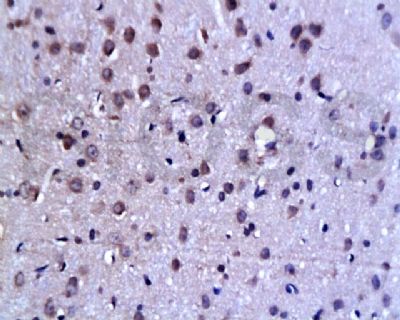
[IF=1.26] Liu et al. Therapeutic effects of 1,25-dihydroxyvitamin D3 on diabetes-induced liver complications in a rat model. (2016) Exp.Ther.Me. 11:2284-2292 IHC,IHSLCP ; Rat.
[IF=1.56] Du, Jinghua, et al. "TLR4‑dependent signaling pathway modulation: A novel mechanism by which pioglitazone protects against nutritional fibrotic steatohepatitis in mice." Molecular medicine reports 13.3 (2016): 2159-2166. WB ; Mouse.
[IF=2.3] Zhang, Ying, et al. "Overexpression of WNT5B promotes COLO 205 cell migration and invasion through the JNK signaling pathway." Oncology Reports. WB ; Human.
[IF=6.551] Mei Ha. et al. PKCα mediated by the PI3K/Akt-FOXA1 cascade facilitates cypermethrin-induced hyperthyroidism. Sci Total Environ. 2021 Feb;757:143727 WB ; Rat.
[IF=4.259] Wu Z et al. Co-infection of Mycoplasma gallisepticum and Escherichia coli Triggers Inflammatory Injury Involving the IL-17 Signaling Pathway. Front Microbiol. 2019 Nov 15;10:2615. WB ; Chicken.
[IF=3.923] Manabe A et al.Upregulation of transient receptor potential melastatin 6 channel expression by rosiglitazone and all-trans-retinoic acid in erlotinib-treated renal tubular epithelial cells.(2018)J Cell Physiol. ICC ;
[IF=5.58] Zhou, Zhiwei, et al. "microRNA let-7c is essential for the anisomycin-elicited apoptosis in Jurkat T cells by linking JNK1/2 to AP-1/STAT1/STAT3 signaling." Scientific Reports 6 (2016): 24434. WB ; Human.
[IF=2.08] Zhang, Jihong, et al. "Interleukin 18 augments growth ability via NF-κB and p38/ATF2 pathways by targeting cylin B1, cyclin B2, cylin A2, and Bcl-2 in BRL-3A rat liver cells." Gene (2015). WB ; Rat.