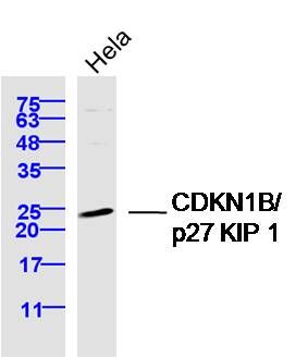
Sample: Hela Cell (Human) Lysate at 40 ug
Primary: Anti-CDKN1B/p27 KIP 1 (SL0742R) at 1/300 dilution
Secondary: IRDye800CW Goat Anti-Rabbit IgG at 1/20000 dilution
Predicted band size: 22 kD
Observed band size: 24 kD
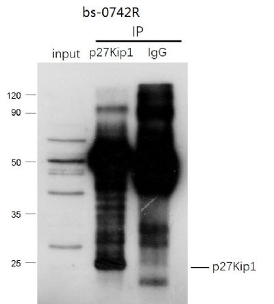
CDKN1B (p27Kip1) was immunoprecipitated from mouse kidney tissue with SL0742R at 1/150 dilution. Western blot was performed from the immunoprecipitate using protein A/G beads. HRP Conjugated Goat anti-Rabbit IgG (Heavy Chain specific) was used as secondary antibody at 1:5000 dilution.
Lane 1: mouse kidney tissue lysate 10 µg (Input).
Lane 2: SL0742R IP in mouse kidney tissue lysate.
Lane 3: native rabbit IgG IP in mouse kidney tissue lysate (negative control).
Secondary
All lanes Goat anti-Rabbit IgG (Heavy Chain specific), HRP Conjugated, 1:5000
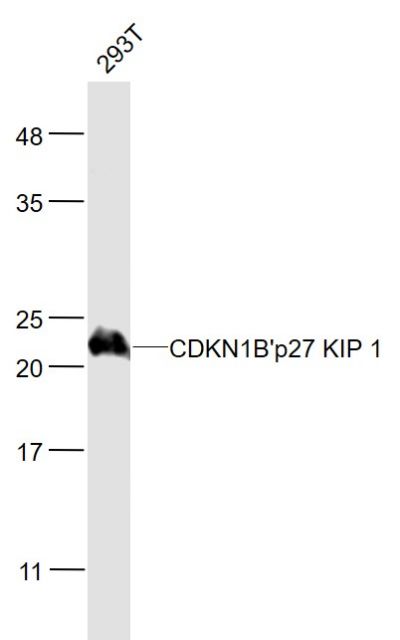
Sample:
293T(Human) Cell Lysate at 30 ug
Primary: Anti- CDKN1B'p27 KIP 1 (SL0742R) at 1/1000 dilution
Secondary: IRDye800CW Goat Anti-Rabbit IgG at 1/20000 dilution
Predicted band size: 22 kD
Observed band size: 22 kD
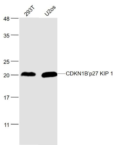
Sample:
293T(Human) Cell Lysate at 30 ug
U2os(Human) Cell Lysate at 30 ug
Primary: Anti- CDKN1B’p27 KIP 1 (SL0742R) at 1/1000 dilution
Secondary: IRDye800CW Goat Anti-Rabbit IgG at 1/20000 dilution
Predicted band size: 22 kD
Observed band size: 22 kD
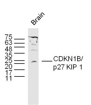
Sample:
Brain (Mouse) Lysate at 40 ug
Primary: Anti- CDKN1B'p27 KIP 1 (SL0742R) at 1/300 dilution
Secondary: IRDye800CW Goat Anti-Rabbit IgG at 1/20000 dilution
Predicted band size: 22 kD
Observed band size: 22 kD
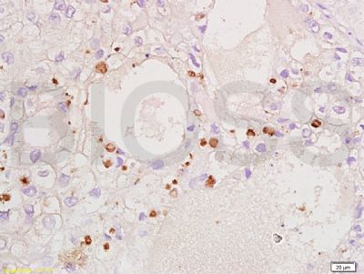
Tissue/cell: human ovary carcinoma; 4% Paraformaldehyde-fixed and paraffin-embedded;
Antigen retrieval: citrate buffer ( 0.01M, pH 6.0 ), Boiling bathing for 15min; Block endogenous peroxidase by 3% Hydrogen peroxide for 30min; Blocking buffer (normal goat serum,SLC0005) at 37∩ for 20 min;
Incubation: Anti-CDKN1B/P27kip1 Polyclonal Antibody, Unconjugated(SL0742R) 1:200, overnight at 4∑C, followed by conjugation to the secondary antibody(SP-0023) and DAB(SLC0010) staining
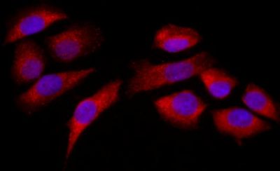
Tissue/cell: MCF7; 4% Paraformaldehyde-fixed; Triton X-100 at room temperature for 20 min; Blocking buffer (normal goat serum, SLC0005) at 37°C for 20 min; Antibody incubation with (CDKN1B ) Polyclonal Antibody, Unconjugated (SL0742R) 1:200, 90 minutes at 37°C; followed by a conjugated Goat Anti-Rabbit IgG antibody (SL0295G-FITC) at 37°C for 90 minutes, DAPI (blue, C02-04002) was used to stain the cell nuclei.
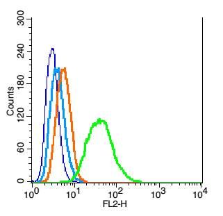
Blank control: RSC96(blue), the cells were fixed with 2% paraformaldehyde (10 min) and then permeabilized with ice-cold 90% methanol for 30 min on ice.
Isotype Control Antibody: Rabbit IgG(orange) ;
Secondary Antibody: Goat anti-rabbit IgG-PE(white blue),
Dilution: 1:200 in 1 X PBS containing 0.5% BSA ;
Primary Antibody Dilution: 1μg in 100μL1X PBS containing 0.5% BSA(green).
|