[IF=15.881] Yuting Shen. et al. Tailoring Chemoimmunostimulant Bioscaffolds for Inhibiting Tumor Growth and Metastasis after Incomplete Microwave Ablation. Acs Nano. 2021;XXXX(XXX):XXX-XXX IF ; Mouse.
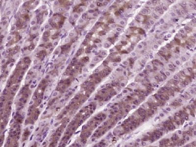
[IF=12.13] Tian Zhang. et al. Supramolecular Tadalafil Nanovaccine for Cancer Immunotherapy by Alleviating Myeloid-Derived Suppressor Cells and Heightening Immunogenicity. 2021 May 13 IHC ; Mouse.
[IF=9.038] Xuting Liu. et al. Amorphous silica nanoparticles induce inflammation via activation of NLRP3 inflammasome and HMGB1/TLR4/MYD88/NF-kb signaling pathway in HUVEC cells. J Hazard Mater. 2021 Feb;404:14850 WB,IF,IP ; Human.
[IF=3.337] Hongzhong Cheng. et al. Circular RNA circLPAR3 Facilitates Esophageal Squamous Cell Carcinoma Progression Through Upregulating HMGB1 via Sponging miR-375/miR-433. Oncotargets Ther. 2020; 13: 7759–7771 WB ; Human.
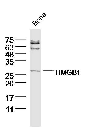
[IF=2.341] Diao S et al. IGF2 enhanced the osteo‐/dentinogenic and neurogenic differentiation potentials of stem cells from apical papilla. J Oral Rehabil. 2019 Jul 10. WB ; Human.
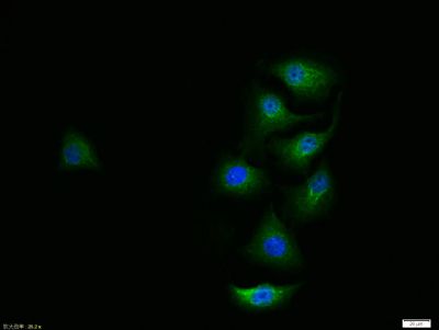
[IF=3.166] Shi Q et al. Polychlorinated biphenyl quinone-induced signaling transition from autophagy to apoptosis is regulated by HMGB1 and p53 in human hepatoma HepG2 cells. Toxicol Lett. 2019 May 15;306:25-34. WB,Co-IP&IF ; Human.
[IF=0] He et al. The effects of n-3 PUFA and intestinal lymph drainage on high-mobility group box 1 and Toll-like receptor 4 mRNA in rats with intestinal ischaemia-reperfusion injury. (2012) Br.J.Nut. 108:883-92 WB, IHSLCP ; Rat.
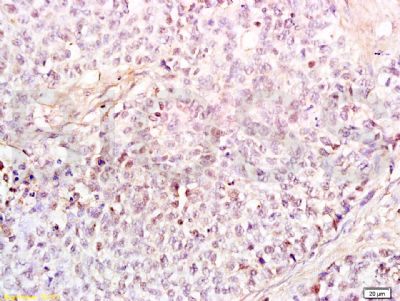
[IF=2.766] Xiang et al. Glycyrrhizin suppresses the expressions of HMGB1 and relieves the severity of traumatic pancreatitis in rats. (2014) PLoS.One. 9:e115982 IHC ; Rat.
[IF=2.61] Li, Yuan-bo, et al. "Methotrexate affects HMGB1 expression in rheumatoid arthritis, and the downregulation of HMGB1 prevents rheumatoid arthritis progression." Molecular and Cellular Biochemistry: 1-10. Human.
[IF=2.35] Li, Guofu, et al. "The neutrophil elastase inhibitor, sivelestat, attenuates sepsis-related kidney injury in rats." International Journal of Molecular Medicine 38.3 (2016): 767-775. WB ; Rat.
[IF=1.34] Sun, Shanping, et al. "High mobility group box-1 and its clinical value in breast cancer." OncoTargets and Therapy 8 (2015): 413. IHSLCP ; Human.
[IF=3.05] Rui, Zhang, et al. "Omega-3 polyunsaturated fatty acids inhibit the increase in cytokines and chemotactic factors induced in vitro by lymph fluid from an intestinal ischemia-reperfusion injury model." Nutrition (2014). WB ; Rat.
[IF=3.24] Zhao, Lu, et al. "The high mobility group box 1 protein of< i> Sciaenops ocellatus is a secreted cytokine that stimulates macrophage activation." Developmental & Comparative Immunology 35.10 (2011): 1052-1058. WB ;
[IF=8.947] Zhen-Han Feng. et al. A combination strategy based on an Au nanorod/doxorubicin gel via mild photothermal therapy combined with antigen-capturing liposomes and anti-PD-L1 agent promote a positive shift in the cancer-immunity cycle. Acta Biomater. 2021 Oct;: IF ; Mouse.
[IF=2.886] Lei Chen. et al. Circ_0032821 Facilitates Gastric Cancer Cell Proliferation, Migration, Invasion and Glycolysis by Regulating MiR-1236-3p/HMGB1 Axis. Cancer Manag Res. 2020; 12: 9965–9976 WB ; Human.
[IF=3.12] Jiang, W., et al. "Curculigoside A attenuates experimental cerebral ischemia injury< i> in vitro and< i> vivo." Neuroscience 192 (2011): 572-579. WB ; Rat.
[IF=6.304] Chen Y et al. Dendritic cells-derived interferon-λ1 ameliorated inflammatory bone destruction through inhibiting osteoclastogenesis. Cell Death Dis. 2020 Jun 2;11(6):414. WB ; Mouse.
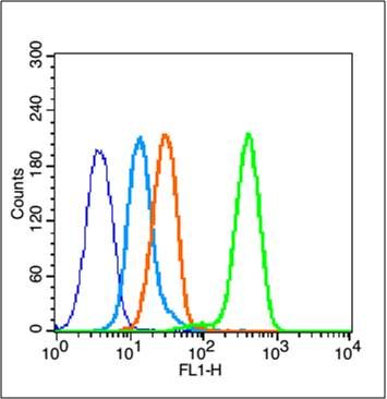
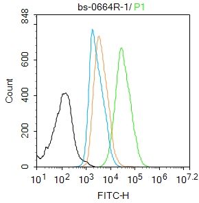
[IF=15.621] Feng B et al. Enhancing Triple Negative Breast Cancer Immunotherapy by ICG‐Templated Self‐Assembly of Paclitaxel Nanoparticles. Advanced Functional Materials,2019 1906605. FCM&ICC ; Mouse.
[IF=3.687] Yuan FH et al. microRNA‐30a inhibits the liver cell proliferation and promotes cell apoptosis through the JAK/STAT signaling pathway by targeting SOCS‐1 in rats with sepsis. J Cell Physiol. 2019 Apr 10. WB ; Rat.
[IF=3.457] Ling L et al. MicroRNA-30e promotes hepatocyte proliferation and inhibits apoptosis in cecal ligation and puncture-induced sepsis through the JAK/STAT signaling pathway by binding to FOSL2.Biomed Pharmacother. 2018 Aug;104:411-419. WB ; Rat.
[IF=1.075] Qiao et al. Effect of different 1, 25-(OH)2D3 doses on high mobility group box1 and toll-like receptors 4 expression in lung tissue of asthmatic mice. (2015) Int.J.Clin.Exp.Med. 8:4016-23 IHC ; Mouse.
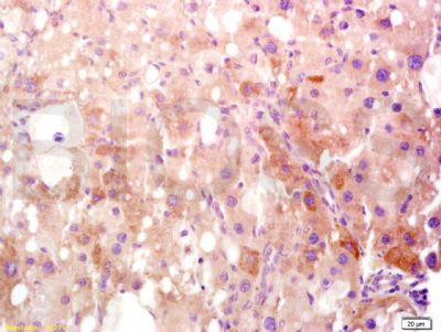
[IF=3.549] Hong et al. Luteolin Treatment Protects against Renal Ischemia-Reperfusion Injury in Rats. (2018) Mediators.Inflamm. 2017:9783893 IHC ; Rat.
[IF=1.89] Sun et al. Expression and Significance of High-Mobility Group Protein B1 (HMGB1) and the Receptor for Advanced Glycation End-Product (RAGE) in Knee Osteoarthritis. (2016) Med.Sci.Monit. 22:2105-12 IHC,WB ; Human.
[IF=0.94] Wang, Xin‑Jun, et al. "Clinical and prognostic significance of high-mobility group box-1 in human gliomas." Experimental and Therapeutic Medicine. WB ; Human.