Home > Product > Antibody > Rabbit Anti-MDH1 antibody
Cytosolic malate dehydrogenase; Malate dehydrogenase 1, NAD (soluble); MDHA; MDHs; Soluble malate dehydrogenase; Malate dehydrogenase; Diiodophenylpyruvate reductase; Malate dehydrogenase 1; Malate dehydrogenase 1 NAD (soluble); Malate dehydrogenase 2; Ma
Cat:
SL3996R
Species Reactivity:
Human,Mouse,Rat,(predicted: Chicken,Dog,Pig,Cow,Horse,Rabbit,)
Immunogen:
KLH conjugated synthetic peptide derived from human MDH1:265-334/334
Format:
Liquid
Storage instructions:
Shipped at 4℃. Store at -20 °C for one year. Avoid repeated freeze/thaw cycles.
Concentration:
1mg/ml
Clonality:
Polyclonal
Isotype:
IgG
Applications:
WB=1:500-2000ELISA=1:5000-10000IHC-P=1:100-500IHC-F=1:100-500Flow-Cyt=0.2ug/TestIF=1:100-500(Paraffin sections need to do antigen repair)not yet tested in other applications.optimal dilutions/concentrations should be determined by the end user.
Host:
Rabbit
Product Overview:
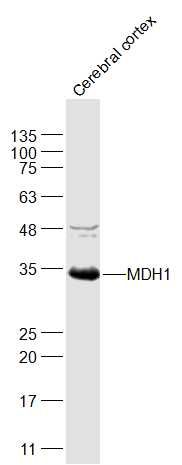
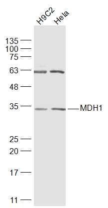
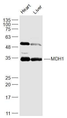
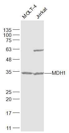
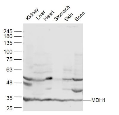
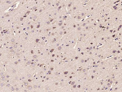
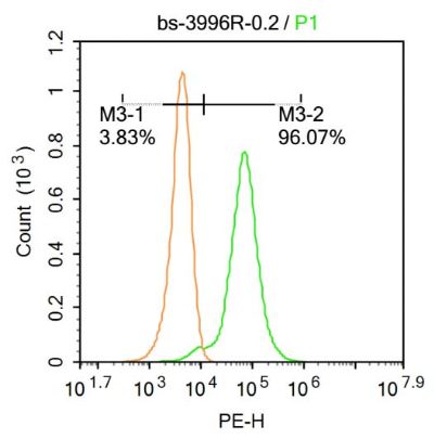
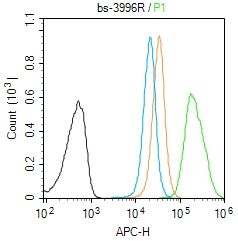
Sample: Cerebral cortex(Rat) Lysate at 40 ugPrimary: Anti-MDH1  (SL3996R) at 1/1000 dilutionSecondary: IRDye800CW Goat Anti-Rabbit IgG at 1/20000 dilutionPredicted band size: 36 kDObserved band size: 34 kDSample: H9C2(Rat) Cell Lysate at 30 ugHela(Human) Cell Lysate at 30 ugPrimary: Anti-MDH1  (SL3996R) at 1/1000 dilutionSecondary: IRDye800CW Goat Anti-Rabbit IgG at 1/20000 dilutionPredicted band size: 36 kDObserved band size: 34 kDSample: Heart (Mouse) Lysate at 40 ugLiver (Mouse) Lysate at 40 ugPrimary: Anti-MDH1  (SL3996R) at 1/1000 dilutionSecondary: IRDye800CW Goat Anti-Rabbit IgG at 1/20000 dilutionPredicted band size: 36 kDObserved band size: 34 kDSample: MOLT-4(Human) Cell Lysate at 30 ugJurkat(Human) Cell Lysate at 30 ugPrimary: Anti-MDH1  (SL3996R) at 1/1000 dilutionSecondary: IRDye800CW Goat Anti-Rabbit IgG at 1/20000 dilutionPredicted band size: 36 kDObserved band size: 34 kDSample: Kidney (Mouse) Lysate at 40 ugLiver (Mouse) Lysate at 40 ugHeart (Mouse) Lysate at 40 ugStomach (Mouse) Lysate at 40 ugSkin (Mouse) Lysate at 40 ugBone (Mouse) Lysate at 40 ugPrimary: Anti- MDH1 (SL3996R) at 1/300 dilutionSecondary: IRDye800CW Goat Anti-Rabbit IgG at 1/20000 dilutionPredicted band size: 36 kDObserved band size: 36 kD Paraformaldehyde-fixed, paraffin embedded (Rat brain); Antigen retrieval by microwave in sodium citrate buffer (pH6.0) ; Block endogenous peroxidase by 3% hydrogen peroxide for 30 minutes; Blocking buffer (3% BSA) at RT for 30min; Antibody incubation with (MDH1) Polyclonal Antibody, Unconjugated (SL3996R) at 1:400 overnight at 4°C, followed by conjugation to the secondary antibody (labeled with HRP)and DAB staining.Blank control:Molt-4. Primary Antibody (green line): Rabbit Anti-MDH1 antibody (SL3996) Dilution: 0.2μg /10^6 cells; Isotype Control Antibody (orange line): Rabbit IgG . Secondary Antibody : Goat anti-rabbit IgG-PEDilution: 0.2μg /test. ProtocolThe cells were fixed with 4% PFA (10min at room temperature)and then permeabilized with 0.1% PBST for 20 min at room temperature. The cells were then incubated in 5%BSA to block non-specific protein-protein interactions for 30 min at at room temperature .Cells stained with Primary Antibody for 30 min at room temperature. The secondary antibody used for 40 min at room temperature. Acquisition of 20,000 events was performed.Blank control (Black line): Molt4 (Black). Primary Antibody (green line):Rabbit Anti-DMT1 antibody (SL3996R)Dilution: 1μg /10^6 cells; Isotype Control Antibody (orange line): Rabbit IgG . Secondary Antibody (white blue line): Goat anti-rabbit IgG-AF647Dilution: 1μg /test. ProtocolThe cells were fixed with 4% PFA (10min at room temperature)and then permeabilized with PBST for 20 min at room temperature. The cells were then incubated in 5%BSA to block non-specific protein-protein interactions for 30 min at room temperature .Cells stained with Primary Antibody for 30 min at room temperature. The secondary antibody used for 40 min at room temperature. Acquisition of 20,000 events was performed.
More
Unit:
Price: $
Product PDFs
Datasheet:


Malate dehydrogenase catalyzes the reversible oxidation of malate to oxaloacetate, utilizing the NAD/NADH cofactor system in the citric acid cycle. Malate dehydrogenase 1 (MDH1) is localized to the cytoplasm and may play pivotal roles in the malate-aspartate shuttle that operates in the metabolic coordination between cytosol and mitochondria.

Subunit:
Homodimer.

Subcellular Location:
Cytoplasm.

Post-translational modifications:
ISGylated.
Acetylation at Lys-118 dramatically enhances enzymatic activity and promotes adipogenic differentiation.

Similarity:
Belongs to the LDH/MDH superfamily. MDH type 2 family.

SWISS:
P40925

Gene ID:
4190

Database links:

Entrez Gene: 4190 Human

Entrez Gene: 17449 Mouse

Entrez Gene: 24551 Rat

Omim: 154200 Human

SwissProt: P40925 Human

SwissProt: P14152 Mouse

SwissProt: O88989 Rat

Unigene: 526521 Human

Unigene: 212703 Mouse

Unigene: 13492 Rat


Picture

Sample:
Cerebral cortex(Rat) Lysate at 40 ug
Primary: Anti-MDH1  (SL3996R) at 1/1000 dilution
Secondary: IRDye800CW Goat Anti-Rabbit IgG at 1/20000 dilution
Predicted band size: 36 kD
Observed band size: 34 kD
Sample:
H9C2(Rat) Cell Lysate at 30 ug
Hela(Human) Cell Lysate at 30 ug
Primary: Anti-MDH1  (SL3996R) at 1/1000 dilution
Secondary: IRDye800CW Goat Anti-Rabbit IgG at 1/20000 dilution
Predicted band size: 36 kD
Observed band size: 34 kD
Sample:
Heart (Mouse) Lysate at 40 ug
Liver (Mouse) Lysate at 40 ug
Primary: Anti-MDH1  (SL3996R) at 1/1000 dilution
Secondary: IRDye800CW Goat Anti-Rabbit IgG at 1/20000 dilution
Predicted band size: 36 kD
Observed band size: 34 kD
Sample:
MOLT-4(Human) Cell Lysate at 30 ug
Jurkat(Human) Cell Lysate at 30 ug
Primary: Anti-MDH1  (SL3996R) at 1/1000 dilution
Secondary: IRDye800CW Goat Anti-Rabbit IgG at 1/20000 dilution
Predicted band size: 36 kD
Observed band size: 34 kD
Sample:
Kidney (Mouse) Lysate at 40 ug
Liver (Mouse) Lysate at 40 ug
Heart (Mouse) Lysate at 40 ug
Stomach (Mouse) Lysate at 40 ug
Skin (Mouse) Lysate at 40 ug
Bone (Mouse) Lysate at 40 ug
Primary: Anti- MDH1 (SL3996R) at 1/300 dilution
Secondary: IRDye800CW Goat Anti-Rabbit IgG at 1/20000 dilution
Predicted band size: 36 kD
Observed band size: 36 kD
Paraformaldehyde-fixed, paraffin embedded (Rat brain); Antigen retrieval by microwave in sodium citrate buffer (pH6.0) ; Block endogenous peroxidase by 3% hydrogen peroxide for 30 minutes; Blocking buffer (3% BSA) at RT for 30min; Antibody incubation with (MDH1) Polyclonal Antibody, Unconjugated (SL3996R) at 1:400 overnight at 4°C, followed by conjugation to the secondary antibody (labeled with HRP)and DAB staining.
Blank control:Molt-4.
Primary Antibody (green line): Rabbit Anti-MDH1 antibody (SL3996)
Dilution: 0.2μg /10^6 cells;
Isotype Control Antibody (orange line): Rabbit IgG .
Secondary Antibody : Goat anti-rabbit IgG-PE
Dilution: 0.2μg /test.
Protocol
The cells were fixed with 4% PFA (10min at room temperature)and then permeabilized with 0.1% PBST for 20 min at room temperature. The cells were then incubated in 5%BSA to block non-specific protein-protein interactions for 30 min at at room temperature .Cells stained with Primary Antibody for 30 min at room temperature. The secondary antibody used for 40 min at room temperature. Acquisition of 20,000 events was performed.
Blank control (Black line): Molt4 (Black).
Primary Antibody (green line):Rabbit Anti-DMT1 antibody (SL3996R)
Dilution: 1μg /10^6 cells;
Isotype Control Antibody (orange line): Rabbit IgG .
Secondary Antibody (white blue line): Goat anti-rabbit IgG-AF647
Dilution: 1μg /test.
Protocol
The cells were fixed with 4% PFA (10min at room temperature)and then permeabilized with PBST for 20 min at room temperature. The cells were then incubated in 5%BSA to block non-specific protein-protein interactions for 30 min at room temperature .Cells stained with Primary Antibody for 30 min at room temperature. The secondary antibody used for 40 min at room temperature. Acquisition of 20,000 events was performed.
Product Feedback Wall
Message :
Your Email :
Copyright © 2007-2018 Sunlong Medical All Rights Reserved.