Home > Product > Antibody > Rabbit Anti-FOXP3 antibody
Forkhead box protein P3; forkhead box P3; Scurfin; Forkhead box protein P3, C-terminally processed; Forkhead box protein P3 41 kDa form; JM2; AIID; IPEX; PIDX; XPID; DIETER; FOXP3_HUMAN;
Cat:
SL10211R
Species Reactivity:
Human,Mouse,Rat,(predicted: Dog,Pig,Cow,Horse,Rabbit,Sheep,Guinea Pig,)
Immunogen:
KLH conjugated synthetic peptide derived from human FoxP3:331-431/431
Format:
Liquid
Storage instructions:
Shipped at 4℃. Store at -20 °C for one year. Avoid repeated freeze/thaw cycles.
Concentration:
1mg/ml
Clonality:
Polyclonal
Isotype:
IgG
Applications:
WB=1:500-2000ELISA=1:5000-10000IHC-P=1:100-500IHC-F=1:100-500Flow-Cyt=0.2ug/TestICC=1:100IF=1:100-500(Paraffin sections need to do antigen repair)not yet tested in other applications.optimal dilutions/concentrations should be determined by the end user.
Host:
Rabbit
Product Overview:
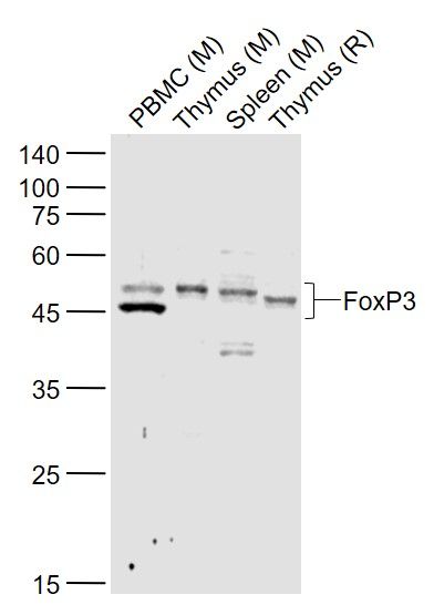
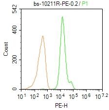
Sample: Lane 1: PBMC (Mouse) Lysate at 40 ugLane 2: Thymus (Mouse) Lysate at 40 ugLane 3: Spleen (Mouse) Lysate at 40 ugLane 4: Thymus (Rat) Lysate at 40 ugPrimary: Anti-FoxP3 (SL10211R) at 1/1000 dilutionSecondary: IRDye800CW Goat Anti-Rabbit IgG at 1/20000 dilutionPredicted band size: 43/45 kDObserved band size: 43/45 kDBlank control: Mouse spleen. Primary Antibody (green line): Rabbit Anti-FoxP3/PE Conjugated antibody (SL10211R-PE) Dilution: 0.2μg /10^6 cells; Isotype Control Antibody (orange line): Rabbit IgG-PE . ProtocolThe cells were fixed with 4% PFA (10min at room temperature)and then permeabilized with 90% ice-cold methanol for 20 min at-20℃. The cells were then incubated in 5% BSA to block non-specific protein-protein interactions for 30 min at room temperature. The cells were stained with Primary Antibody for 30 min at room temperature. Acquisition of 20,000 events was performed.Blank control: MCF7. Primary Antibody (green line): Rabbit Anti-FoxP3 antibody (SL10211R) Dilution: 2μg /10^6 cells; Isotype Control Antibody (orange line): Rabbit IgG . Secondary Antibody : Goat anti-rabbit IgG-AF647Dilution: 1μg /test. ProtocolThe cells were fixed with 4% PFA (10min at room temperature)and then permeabilized with 90% ice-cold methanol for 20 min at-20℃.The cells were then incubated in 5%BSA to block non-specific protein-protein interactions for 30 min at room temperature .Cells stained with Primary Antibody for 30 min at room temperature. The secondary antibody used for 40 min at room temperature. Acquisition of 20,000 events was performed.Blank control: MCF7. Primary Antibody (green line): Rabbit Anti-FoxP3 antibody (SL10211R) Dilution: 2μg /10^6 cells; Isotype Control Antibody (orange line): Rabbit IgG . Secondary Antibody : Goat anti-rabbit IgG-AF647Dilution: 1μg /test. ProtocolThe cells were fixed with 4% PFA (10min at room temperature)and then permeabilized with 90% ice-cold methanol for 20 min at-20℃.The cells were then incubated in 5%BSA to block non-specific protein-protein interactions for 30 min at room temperature .Cells stained with Primary Antibody for 30 min at room temperature. The secondary antibody used for 40 min at room temperature. Acquisition of 20,000 events was performed.Blank control (Black line):Mouse spleen (Black). Primary Antibody (green line): Rabbit Anti-FoxP3 antibody (SL10211R) Dilution: 3μg /10^6 cells; Isotype Control Antibody (orange line): Rabbit IgG . Secondary Antibody (white blue line): Goat anti-rabbit IgG-PEDilution: 1μg /test. ProtocolThe cells were fixed with 4% PFA (10min at room temperature)and then permeabilized with 90% ice-cold methanol for 20 min at room temperature. The cells were then incubated in 5%BSA goat serum to block non-specific protein-protein interactions for 15 min at room temperature .Cells stained with Primary Antibody for 30 min at room temperature. The secondary antibody used for 40 min at room temperature. Acquisition of 20,000 events was performed.
More
Unit:
Price: $
Product PDFs
Datasheet:


The protein encoded by this gene is a member of the forkhead/winged-helix family of transcriptional regulators. Defects in this gene are the cause of immunodeficiency polyendocrinopathy, enteropathy, X-linked syndrome (IPEX), also known as X-linked autoimmunity-immunodeficiency syndrome. Alternatively spliced transcript variants encoding different isoforms have been identified. [provided by RefSeq, Jul 2008].

Function:
Probable transcription factor. Plays a critical role in the control of immune response.

Subunit:
Interacts with IKZF3.

Subcellular Location:
Nucleus (Potential).

Post-translational modifications:
Acetylation on lysine residues stabilizes FOXP3 and promotes differentiation of T-cells into induced regulatory T-cells (iTregs) associated with suppressive functions. Deacetylated by SIRT1.

DISEASE:
Defects in FOXP3 are the cause of immunodeficiency polyendocrinopathy, enteropathy, X-linked syndrome (IPEX) [MIM:304790]; also known as X-linked autoimmunity-immunodeficiency syndrome. IPEX is characterized by neonatal onset insulin-dependent diabetes mellitus, infections, secretory diarrhea, trombocytopenia, anemia and eczema. It is usually lethal in infancy.

Similarity:
Contains 1 C2H2-type zinc finger.
Contains 1 fork-head DNA-binding domain.

SWISS:
Q9BZS1

Gene ID:
50943

Database links:

Entrez Gene: 50943 Human

Entrez Gene: 20371 Mouse

Entrez Gene: 317382 Rat

Omim: 300292 Human

SwissProt: Q9BZS1 Human

SwissProt: Q99JB6 Mouse

SwissProt: D3ZKI1 Rat

Unigene: 247700 Human

Unigene: 182291 Mouse


Picture

Sample:
Lane 1: PBMC (Mouse) Lysate at 40 ug
Lane 2: Thymus (Mouse) Lysate at 40 ug
Lane 3: Spleen (Mouse) Lysate at 40 ug
Lane 4: Thymus (Rat) Lysate at 40 ug
Primary: Anti-FoxP3 (SL10211R) at 1/1000 dilution
Secondary: IRDye800CW Goat Anti-Rabbit IgG at 1/20000 dilution
Predicted band size: 43/45 kD
Observed band size: 43/45 kD
Blank control: Mouse spleen.
Primary Antibody (green line): Rabbit Anti-FoxP3/PE Conjugated antibody (SL10211R-PE)
Dilution: 0.2μg /10^6 cells;
Isotype Control Antibody (orange line): Rabbit IgG-PE .
Protocol
The cells were fixed with 4% PFA (10min at room temperature)and then permeabilized with 90% ice-cold methanol for 20 min at-20℃. The cells were then incubated in 5% BSA to block non-specific protein-protein interactions for 30 min at room temperature. The cells were stained with Primary Antibody for 30 min at room temperature. Acquisition of 20,000 events was performed.
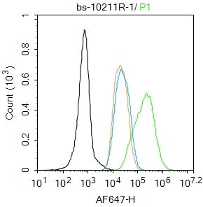
Blank control: MCF7.
Primary Antibody (green line): Rabbit Anti-FoxP3 antibody (SL10211R)
Dilution: 2μg /10^6 cells;
Isotype Control Antibody (orange line): Rabbit IgG .
Secondary Antibody : Goat anti-rabbit IgG-AF647
Dilution: 1μg /test.
Protocol
The cells were fixed with 4% PFA (10min at room temperature)and then permeabilized with 90% ice-cold methanol for 20 min at-20℃.The cells were then incubated in 5%BSA to block non-specific protein-protein interactions for 30 min at room temperature .Cells stained with Primary Antibody for 30 min at room temperature. The secondary antibody used for 40 min at room temperature. Acquisition of 20,000 events was performed.
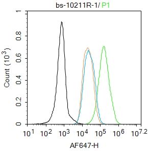
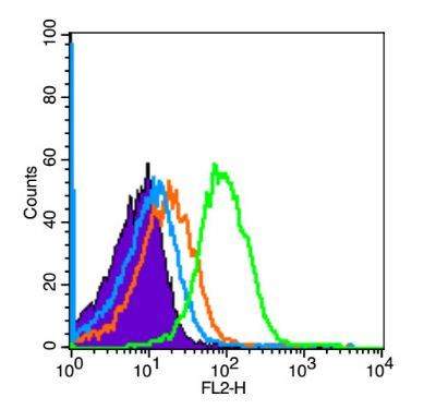
Blank control (Black line):Mouse spleen (Black).
Primary Antibody (green line): Rabbit Anti-FoxP3 antibody (SL10211R)
Dilution: 3μg /10^6 cells;
Isotype Control Antibody (orange line): Rabbit IgG .
Secondary Antibody (white blue line): Goat anti-rabbit IgG-PE
Dilution: 1μg /test.
Protocol
The cells were fixed with 4% PFA (10min at room temperature)and then permeabilized with 90% ice-cold methanol for 20 min at room temperature. The cells were then incubated in 5%BSA goat serum to block non-specific protein-protein interactions for 15 min at room temperature .Cells stained with Primary Antibody for 30 min at room temperature. The secondary antibody used for 40 min at room temperature. Acquisition of 20,000 events was performed.
Product Feedback Wall
Message :
Your Email :
Copyright © 2007-2018 Sunlong Medical All Rights Reserved.