[IF=2.217] Qi Wang. et al. Effect of oral administration of Limosilactobacillus reuteri on intestinal barrier function and mucosal immunity of suckling piglets. Ital J Anim Sci. 2022;21(1):612-623 WB ; Piglets.
[IF=3.414] Hu N et al. Phillygenin inhibits LPS-induced activation and inflammation of LX2 cells by TLR4/MyD88/NF-κB signaling pathway. J Ethnopharmacol. 2019 Nov 1:112361. WB ; Human.
[IF=5.717] Na Chen. et al. Casein Oligochitosan-Glycation by Transglutaminase Enhances the Anti-Inflammatory Potential of Casein Hydrolysates to the Lipopolysaccharide-Stimulated IESLC6 Cells. Nutrients. 2022 Jan;14(3):686 WB ; Rat.
[IF=6.419] Quan Rao. et al. Dendritic cell combination therapy reduces the toxicity of triptolide and ameliorates colitis in murine models. Drug Deliv. 2022;29(1):679-691 WB ; Mouse.
[IF=5.246] Zou Xiong. et al. Toll-Like Receptors Serve as Biomarkers for Early Diagnosis and Prognosis Assessment of Kidney Renal Clear Cell Carcinoma by Influencing the Immune Microenvironment: Comprehensive Bioinformatics Analysis Combined With Experimental Validation. Front Mol Biosci. 2022 Jan;0:24 FC ; Human.
[IF=4.411] Shi-Qing Cai. et al. The In Vitro Anti-Inflammatory Activities of Galangin and Quercetin towards the LPS-Injured Rat Intestinal Epithelial (IESLC6) Cells as Affected by Heat Treatment. Molecules. 2021 Jan;26(24):7495 WB ; Rat.
[IF=5.81] Xu X. et al. Alhagi pseudalhagi Extract Exerts Protective Effects Against Intestinal Inflammation in Ulcerative Colitis by Affecting TLR4-Dependent NF-κB Signaling Pathways.. Front Pharmacol. 2021 Nov;12:76922-76922 WB ; Mouse.
[IF=5.878] Chiara Corpetti. et al. Cannabidiol inhibits SARS-Cov-2 spike (S) protein-induced cytotoxicity and inflammation through a PPARγ-dependent TLR4/NLRP3/Caspase-1 signaling suppression in Caco-2 cell line. 2021 Oct 12 WB ; Human.
[IF=2.1] Chen W et al. Integrated analysis of a lncRNA‑mRNA network reveals a potential mechanism underlying necrotizing enterocolitis. Mol Med Rep
. 2020 Jul;22(1):423-435. WB ; human.
[IF=3.845] Peng LY et al. Protective Effect of Piceatannol Against Acute Lung Injury Through Protecting the Integrity of Air-Blood Barrier and Modulating the TLR4/NF-κB Signaling Pathway Activation. Front Pharmacol. 2020 Jan 22;10:1613. WB ; Mouse.
[IF=2.553] Liu W et al. Suppressive effect of glycyrrhizic acid against lipopolysaccharide-induced neuroinflammation and cognitive impairment in C57 mice via toll-like receptor 4 signaling pathway. Food Nutr Res. 2019 Apr 29;63. IHC ; Mouse.
[IF=3.687] Yuan FH et al. microRNA‐30a inhibits the liver cell proliferation and promotes cell apoptosis through the JAK/STAT signaling pathway by targeting SOCS‐1 in rats with sepsis. J Cell Physiol. 2019 Apr 10. WB ; Rat.
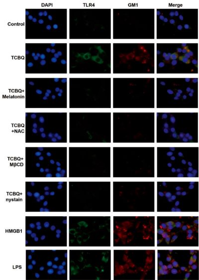
[IF=3.432] Dong W et al. Polychlorinated biphenyl quinone induces caspase 1-mediated pyroptosis through the induction of pro-inflammatory HMGB1-TLR4-NLRP3-GSDMD signal axis. Chem Res Toxicol. 2019 Apr 23. WB&ICF ; Human.
[IF=2.276] Zhao Yonglin. et al. Inhibition of Macrophage Migration Inhibitory Factor Protects against Inflammation through a Toll-like Receptor-Related Pathway after Diffuse Axonal Injury in Rats. Biomed Res Int. 2020;2020:5946205 WB ; Rat.
[IF=2.984] Yitong Pan. et al. N-acetyl-L-tryptophan attenuates hepatic ischemia-reperfusion injury via regulating TLR4/NLRP3 signaling pathway in rats. Peerj. 2021 Aug;9:e11909 WB,IF ; Rat.
[IF=4.932] Alessandro Del Re. et al. Ultramicronized Palmitoylethanolamide Inhibits NLRP3 Inflammasome Expression and Pro-Inflammatory Response Activated by SARS-CoSLV2 Spike Protein in Cultured Murine Alveolar Macrophages. Metabolites. 2021 Sep;11(9):592 WB,IF ; mouse.
[IF=4.451] Meilan Xue. et al. Neuroprotective effect of fucoidan by regulating gut-microbiota-brain axis in alcohol withdrawal mice. J Funct Foods. 2021 Nov;86:104726 WB ; mouse.
[IF=4.162] Jiali Yang. et al. Baicalin Rescues Cognitive Dysfunction, Mitigates Neurodegeneration, and Exerts Anti-Epileptic Effects Through Activating TLR4/MYD88/Caspase-3 Pathway in Rats. Drug Des Dev Ther. 2021 Jul;15:3163-336 IHC ; Rat.
[IF=4.235] Zhao Xueqin. et al. The Antimicrobial Peptide Mastoparan X Protects Against Enterohemorrhagic Escherichia coli O157:H7 Infection, Inhibits Inflammation, and Enhances the Intestinal Epithelial Barrier. Front Microbiol. 2021 Jun;12:1392 IF ; Pig.
[IF=1.951] Li Liu. et al. Inhibition of inducible nitric oxide synthase improved erectile dysfunction in rats with type 1 diabetes. 2021 Jun 16 WB ; Rat.
[IF=3.37] Xuelian Ma. et al. Down-regulated long non-coding RNA RMST ameliorates dopaminergic neuron damage in Parkinson’s disease rats via regulation of TLR/NF-κB signaling pathway. Brain Res Bull. 2021 Apr;: WB ; Rat.
[IF=4.147] Sijie Yuan. et al. Bacteroides vulgatus diminishes colonic microbiota dysbiosis ameliorating lumbar bone loss in ovariectomized mice. Bone. 2021 Jan;142:115710 WB ; Mouse.
[IF=3.69] Naihua Hu. et al. Forsythiae Fructuse water extract attenuates liver fibrosis via TLR4/MyD88/NF-κB and TGF-β/smads signaling pathways. J Ethnopharmacol. 2020 Nov;262:113275 WB ; Rat.
[IF=1.918] Dongfeng Cai. et al. The Effects of microRNA-515-5p on the Toll-Like Receptor 4 (TLR4)/JNK Signaling Pathway and WNT1-Inducible-Signaling Pathway Protein 1 (WISP-1) Expression in Rheumatoid Arthritis Fibroblast-Like Synovial (RAFLS) Cells Following Treatment with Receptor Act. Med Sci Monitor. 2020; 26: e920611-1–e920611-9 WB ;
[IF=2.215] Karadag Remzi. et al. Effects of Different Doses of Systemic Isotretinoin on Eyes: A Histopathological and Immunohistochemical Study in Rats. Cornea. 2020 May;39(5):621-627 WB ; Human.
[IF=2.1] Wenjuan Chenet al. Integrated analysis of a lncRNA‑mRNA network reveals a potential mechanism underlying necrotizing enterocolitis. Mol Med Rep
. 2020 Jul;22(1):423-435. WB ; Human.
[IF=1.287] Li X et al. Leptin improves intestinal flora dysfunction in mice with high-fat diet-induced obesity. J Int Med Res. 2020 Jun;48(6):300060520920062. WB ; Mouse.
[IF=1.984] Wang X et al. trans-Cinnamaldehyde Reverses Depressive-Like Behaviors in Chronic Unpredictable Mild Stress Rats by Inhibiting NF-κB/NLRP3 Inflammasome Pathway. Evid Based Complement Alternat Med. 2020 Feb 28;2020:4572185. ELISA ; rat.
[IF=3.457] Ling L et al. MicroRNA-30e promotes hepatocyte proliferation and inhibits apoptosis in cecal ligation and puncture-induced sepsis through the JAK/STAT signaling pathway by binding to FOSL2.Biomed Pharmacother. 2018 Aug;104:411-419. WB ; Rat.