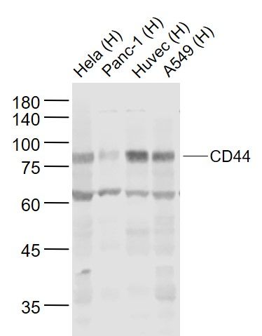
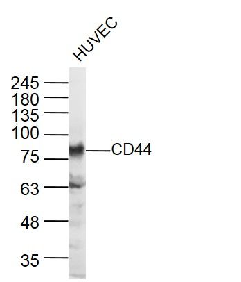
[IF=25.841] Xin Zhou. et al. Tumour-derived extracellular vesicle membrane hybrid lipid nanovesicles enhance siRNA delivery by tumour-homing and intracellular freeway transportation. J Extracell Vesicles. 2022 Mar;11(3):e12198 WB ; Human.
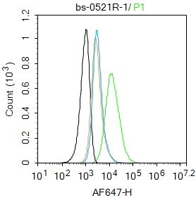
[IF=3.647] Xiaohui Chen. et al. Bone marrow mesenchymal stem cell-derived extracellular vesicles containing miR-497-5p inhibit RSPO2 and accelerate OPLL. Life Sci. 2021 Apr;:119481 FC ; Human.
[IF=8.352] Chanjuan Dong. et al. Graphene-based conductive fibrous scaffold boosts sciatic nerve regeneration and functional recovery upon electrical stimulation. Appl Mater Today. 2020 Dec;21:100870 IHC ; Rat.
[IF=10.317] Kaiyuan Wanget al. An exosome-like programmable-bioactivating paclitaxel prodrug nanoplatform for enhanced breast cancer metastasis inhibition. Biomaterials
. 2020 Oct;257:120224. WB ; Human.
[IF=4.3] Xiong et al. Bone Marrow Mesenchymal Stem-Cell Transplantation Promotes Functional Improvement Associated with CNTF-STAT3 Activation after Hemi-Sectioned Spinal Cord Injury in Tree Shrews. (2017) Front.Cell.Neurosci. 11:172 tree shrews.
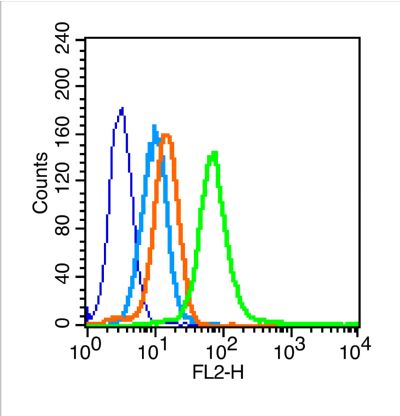
[IF=1.26] Liao et al. Bone mesenchymal stem cells co-expressing VEGF and BMP-6 genes to combat avascular necrosis of the femoral head. (2018) Exp.Ther.Med. 15:954-962 FCM ; Rat.
[IF=5.1] Neal et al. The glycoprotein GPNMB attenuates astrocyte inflammatory responses through the CD44 receptor. (2018) J.Neuroinflammation. 15:73 IF(ICC) ; Mouse.
[IF=4.175] Yue Wu. et al. LncRNA WDFY3-AS2 promotes cisplatin resistance and the cancer stem cell in ovarian cancer by regulating hsa-miR-139-5p/SDC4 axis. Cancer Cell Int. 2021 Dec;21(1):1-14 FC ; Human.
[IF=2.626] Zhao, Gang. et al. LINC02381, a sponge of miR-21, weakens osteogenic differentiation of hUSLCMSCs through KLF12-mediated Wnt4 transcriptional repression. 2021 Nov 15 WB ; Mouse,Human.
[IF=7.727] Xue Yanget al. “Star” miR-34a and CXCR4 antagonist based nanoplex for binary cooperative migration treatment against metastatic breast cancer. J Control Release
. 2020 Oct 10;326:615-627. WB ; Human.
[IF=0.918] Chen F et al.
The biological characteristics of sheep umbilical cord mesenchymal stem cells.Can J Vet Res. 2018 Jul;82(3):216-224. ICF ; lamb.
[IF=1.461] Pei W et al. Biological characterization and pluripotent identification of ovine amniotic fluid stem cells. Cytotechnology. 2018 Jun;70(3):1009-1021. IF&FCM ; ovine embryo.
[IF=1.69] Xu et al. NIBP impacts on the expression of E-cadherin, CD44 and vimentin in colon cancer via the NF-κB pathway. (2016) Mol.Med.Rep. 13:5379-85 WB ; Human.
[IF=1.56] Wang, Xuming, et al. "Characterization of sphere‑forming cells with stem‑like properties from the gastric cancer cell lines MKN45 and SGC7901." Molecular Medicine Reports 10.6 (2014): 2937-2941. FCM ; Human.