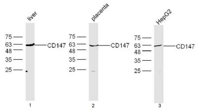
Sample:
Liver(Mouse) Lysate at 40 ug
Placenta(Mouse) Lysate at 40 ug
HepG2 Cell Lysate at 40 ug
Primary: Anti-CD147 (SL0684R) at 1/300 dilution
�
Secondary: IRDye800CW Goat Anti-Rabbit IgG at 1/20000 dilution
�
Predicted band size: 40 kD
Observed band size: 60 kD
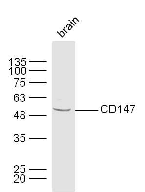
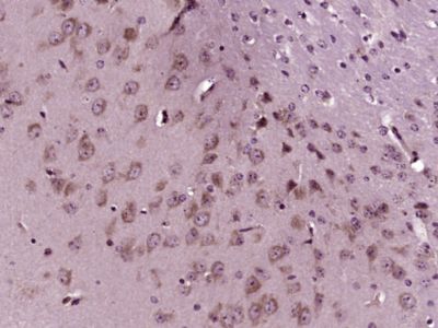
Sample:
Brain (Mouse) Lysate at 40 ug
Primary: Anti-CD147 (Bs-0684R) at 1/300 dilution
Secondary: IRDye800CW Goat Anti-Rabbit IgG at 1/20000 dilution
Predicted band size: 40 kD
Observed band size: 50 kD
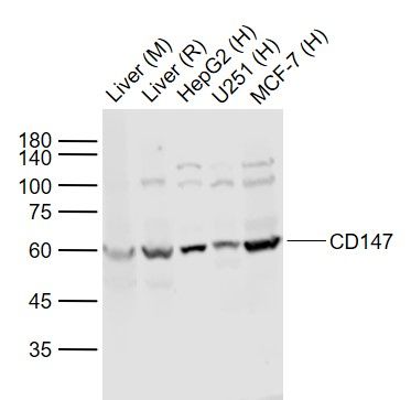
Sample:
Lane 1: Liver (Mouse) Lysate at 40 ug
Lane 2: Liver (Rat) Lysate at 40 ug
Lane 3: HepG2 (Human) Cell Lysate at 30 ug
Lane 4: U251 (Human) Cell Lysate at 30 ug
Lane 5: MCF-7 (Human) Cell Lysate at 30 ug
Primary: Anti-CD147 (SL0684R) at 1/1000 dilution
Secondary: IRDye800CW Goat Anti-Rabbit IgG at 1/20000 dilution
Predicted band size: 50-60 kD
Observed band size: 60 kD
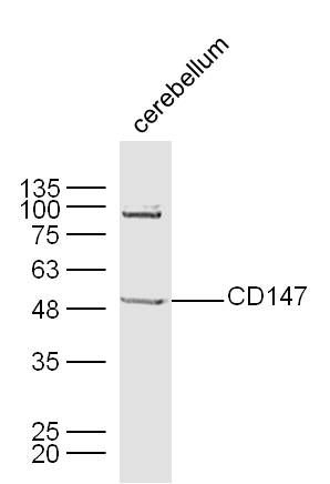
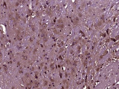
Sample:
Cerebellum (Mouse) Lysate at 40 ug
Primary: Anti-CD147 (Bs-0684R) at 1/300 dilution
Secondary: IRDye800CW Goat Anti-Rabbit IgG at 1/20000 dilution
Predicted band size: 40 kD
Observed band size: 50 kD
Paraformaldehyde-fixed, paraffin embedded (Mouse cerebellum); Antigen retrieval by boiling in sodium citrate buffer (pH6.0) for 15min; Block endogenous peroxidase by 3% hydrogen peroxide for 20 minutes; Blocking buffer (normal goat serum) at 37°C for 30min; Antibody incubation with (CD147) Polyclonal Antibody, Unconjugated (SL0684R) at 1:400 overnight at 4°C, followed by operating according to SP Kit(Rabbit) (sp-0023) instructionsand DAB staining.
Paraformaldehyde-fixed, paraffin embedded (Mouse brain); Antigen retrieval by boiling in sodium citrate buffer (pH6.0) for 15min; Block endogenous peroxidase by 3% hydrogen peroxide for 20 minutes; Blocking buffer (normal goat serum) at 37°C for 30min; Antibody incubation with (CD147) Polyclonal Antibody, Unconjugated (SL0684R) at 1:400 overnight at 4°C, followed by operating according to SP Kit(Rabbit) (sp-0023) instructionsand DAB staining.
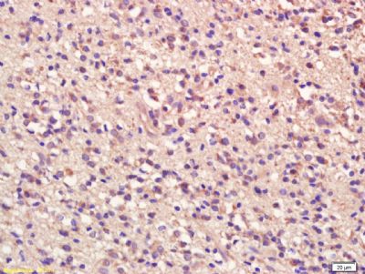
Tissue/cell: human glioma tissue; 4% Paraformaldehyde-fixed and paraffin-embedded;
Antigen retrieval: citrate buffer ( 0.01M, pH 6.0 ), Boiling bathing for 15min; Block endogenous peroxidase by 3% Hydrogen peroxide for 30min; Blocking buffer (normal goat serum,SLC0005) at 37℃ for 20 min;
Incubation: Anti-CD147/TCSF/Emmprin Polyclonal Antibody, Unconjugated(SL0684R) 1:200, overnight at 4°C, followed by conjugation to the secondary antibody(SP-0023) and DAB(SLC0010) staining
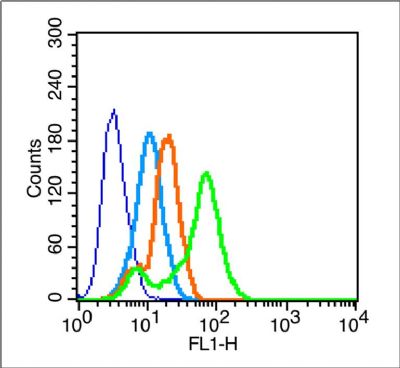
Blank control (blue line): MCF7 (blue).
Primary Antibody (green line):Rabbit Anti-CD147 antibody(SL0684R)
Dilution: 1μg /10^6 cells;
Isotype Control Antibody (orange line): Rabbit IgG .
Secondary Antibody (white blue line): F(ab’)2 fragment goat anti-rabbit IgG-FITC
Dilution: 1μg /test.
Protocol
The cells were fixed with 2% paraformaldehyde for 10 min at room temperature. Cells stained with Primary Antibody for 30 min at room temperature. The cells were then incubated in 1 X PBS/2%BSA/10% goat serum to block non-specific protein-protein interactions followed by the antibody for 15 min at room temperature. The secondary antibody used for 40 min at room temperature. Acquisition of 20,000 events was performed.
|