Home > Product > Antibody > Rabbit Anti-Connexin 43 antibody
Connexin 43; connexin43; Connexin43v; Cx 43v; CX43; CX 43; CX-43; DFNB38; Gap junction 43 kDa heart protein;Connexin-43; Gap junction alpha 1 protein; Gap junction protein alpha 1 43kDa (connexin 43); Gap junction protein alpha 1 43kDa; Gap junction prote
Cat:
SL0651R
Species Reactivity:
Human,Mouse,Rat,Dog,(predicted: Chicken,Cow,)
Immunogen:
KLH conjugated synthetic peptide derived from human Connexin-43:211-260/382 human<Cytoplasmic>
Format:
Liquid
Storage instructions:
Shipped at 4℃. Store at -20 °C for one year. Avoid repeated freeze/thaw cycles.
Concentration:
1mg/ml
Clonality:
Polyclonal
Isotype:
IgG
Applications:
WB=1:500-2000ELISA=1:5000-10000IHC-P=1:100-500IHC-F=1:100-500Flow-Cyt=1μg/TestICC=1:100IF=1:100-500(Paraffin sections need to do antigen repair)not yet tested in other applications.optimal dilutions/concentrations should be determined by the end user.
Host:
Rabbit
Product Overview:
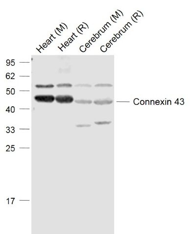
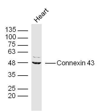
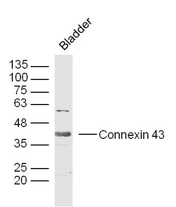
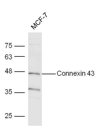
Sample: Lane 1: Heart (Mouse) Lysate at 40 ugLane 2: Heart (Rat) Lysate at 40 ugLane 3: Cerebrum (Mouse) Lysate at 40 ugLane 4: Cerebrum (Rat) Lysate at 40 ugPrimary: Anti-Connexin 43 (SL0651R) at 1/1000 dilutionSecondary: IRDye800CW Goat Anti-Rabbit IgG at 1/20000 dilutionPredicted band size: 37-45 kDObserved band size: 45 kDSample:Heart (Mouse) Lysate at 40 ugPrimary: Anti- Connexin 43(SL0651R)at 1/300 dilutionSecondary: IRDye800CW Goat Anti-Rabbit IgG at 1/20000 dilutionPredicted band size: 42 kDObserved band size: 42/48 kDSample:Bladder (Mouse) Lysate at 40 ugPrimary: Anti- Connexin 43(SL0651R)at 1/300 dilutionSecondary: IRDye800CW Goat Anti-Rabbit IgG at 1/20000 dilutionPredicted band size: 42 kDObserved band size: 42/48 kDSample: Mcf-7 Cell Lysate at 40 ugPrimary: Anti-Connexin(SL0651R) at 1/300 dilutionSecondary: IRDye800CW Goat Anti-Rabbit IgG at 1/10000 dilutionPredicted band size: 42 kDObserved band size: 43 kD Paraformaldehyde-fixed, paraffin embedded (Mouse brain); Antigen retrieval by boiling in sodium citrate buffer (pH6.0) for 15min; Block endogenous peroxidase by 3% hydrogen peroxide for 20 minutes; Blocking buffer (normal goat serum) at 37°C for 30min; Antibody incubation with (Connexin 43) Polyclonal Antibody, Unconjugated (SL0651R) at 1:400 overnight at 4°C, followed by operating according to SP Kit(Rabbit) (sp-0023) instructionsand DAB staining.Paraformaldehyde-fixed, paraffin embedded (Human stomach cancer); Antigen retrieval by boiling in sodium citrate buffer (pH6.0) for 15min; Block endogenous peroxidase by 3% hydrogen peroxide for 20 minutes; Blocking buffer (normal goat serum) at 37°C for 30min; Antibody incubation with (Connexin 43) Polyclonal Antibody, Unconjugated (SL0651R) at 1:200 overnight at 4°C, followed by operating according to SP Kit(Rabbit) (sp-0023) instructions and DAB staining.Tissue/cell:U-251 cell; 4% Paraformaldehyde-fixed; Triton X-100 at room temperature for 20 min; Blocking buffer (normal goat serum, SLC0005) at 37°C for 20 min; Antibody incubation with (Connexin 43) polyclonal Antibody, Unconjugated (SL0651R) 1:100, 90 minutes at 37°C; followed by a FITC conjugated Goat Anti-Rabbit IgG antibody at 37°C for 90 minutes, DAPI (blue, C02-04002) was used to stain the cell nuclei.Tissue/cell: MCF7 cell; 4% Paraformaldehyde-fixed; Triton X-100 at room temperature for 20 min; Blocking buffer (normal goat serum, SLC0005) at 37°C for 20 min; Antibody incubation with (Connexin 43) polyclonal Antibody, Unconjugated (SL0651R) 1:100, 90 minutes at 37°C; followed by a FITC conjugated Goat Anti-Rabbit IgG antibody at 37°C for 90 minutes, DAPI (blue, C02-04002) was used to stain the cell nuclei.Tissue/cell: rat heart tissue;4% Paraformaldehyde-fixed and paraffin-embedded; Antigen retrieval: citrate buffer ( 0.01M, pH 6.0 ), Boiling bathing for 15min; Blocking buffer (normal goat serum,SLC0005) at 37℃ for 20 min; Incubation: Anti-Connexin 43 Polyclonal Antibody, Unconjugated(SL0651R) 1:200, overnight at 4°C; The secondary antibody was Goat Anti-Rabbit IgG, PE conjugated(SL0295G-PE)used at 1:200 dilution for 40 minutes at 37°C. Blank control (blue line):Hela(blue). Primary Antibody (green line): Rabbit Anti-Connexin 43 antibody(SL0651R) Dilution: 1μg /10^6 cells; Isotype Control Antibody (orange line): Rabbit IgG . Secondary Antibody (white blue line): F(ab’)2 fragment goat anti-rabbit IgG-FITC.Dilution: 1μg /test. ProtocolThe cells were fixed with 2% paraformaldehyde (10 min) , then permeabilized with 90% ice-cold methanol for 30 min on ice.Cells stained with Primary Antibody for 30 min at room temperature. The cells were then incubated in 1 X PBS/2%BSA/10% goat serum to block non-specific protein-protein interactions followed by the antibody for 15 min at room temperature. The secondary antibody used for 40 min at room temperature. Acquisition of 20,000 events was performed.
More
Unit:
Price: $
Product PDFs
Datasheet:


This gene is a member of the connexin gene family. The encoded protein is a component of gap junctions, which are composed of arrays of intercellular channels that provide a route for the diffusion of low molecular weight materials from cell to cell. The encoded protein is the major protein of gap junctions in the heart that are thought to have a crucial role in the synchronized contraction of the heart and in embryonic development. A related intronless pseudogene has been mapped to chromosome 5. Mutations in this gene have been associated with oculodentodigital dysplasia and heart malformations. [provided by RefSeq].

Function:
Gap junction protein that acts as a regulator of bladder capacity. A gap junction consists of a cluster of closely packed pairs of transmembrane channels, the connexons, through which materials of low MW diffuse from one cell to a neighboring cell. May play a critical role in the physiology of hearing by participating in the recycling of potassium to the cochlear endolymph. Negative regulator of bladder functional capacity: acts by enhancing intercellular electrical and chemical transmission, thus sensitizing bladder muscles to cholinergic neural stimuli and causing them to contract.

Subunit:
A connexon is composed of a hexamer of connexins. Interacts (via SLCterminus) with TJP1. Interacts (via SLCterminus) with SRC (via SH3 domain). Interacts with UBQLN4. Interacts with SGSM3. Interacts with KIAA1432/CIP150. Interacts with CNST and CSNK1D.

Subcellular Location:
Cell membrane; Multi-pass membrane protein. Cell junction, gap junction.

Tissue Specificity:
Expressed in the heart and fetal cochlea.

Post-translational modifications:
Phosphorylated at Ser-368 by PRKCG; phosphorylation induces disassembly of gap junction plaques and inhibition of gap junction activity. Phosphorylation at Ser-325, Ser-328 and Ser-330 by CK1 modulates gap junction assembly.

DISEASE:
Defects in GJA1 are the cause of autosomal dominant oculodentodigital dysplasia (ODDD) [MIM:164200]; also known as oculodentoosseous dysplasia. ODDD is a highly penetrant syndrome presenting with craniofacial (ocular, nasal, dental) and limb dysmorphisms, spastic paraplegia, and neurodegeneration. Craniofacial anomalies tipically include a thin nose with hypoplastic alae nasi, small anteverted nares, prominent columnella, and microcephaly. Brittle nails and hair abnormalities of hypotrichosis and slow growth are present. Ocular defects include microphthalmia, microcornea, cataracts, glaucoma, and optic atrophy. Syndactyly type 3 and conductive deafness can occur in some cases. Cardiac abnormalities are observed in rare instances.

Similarity:
Belongs to the connexin family. Alpha-type (group II) subfamily.

SWISS:
P17302

Gene ID:
2697

Database links:

Entrez Gene: 2697 Human

Entrez Gene: 1929 Mouse

Entrez Gene: 24392 Rat

Omim: 121014 Human

SwissProt: P17302 Human

SwissProt: P23242 Mouse

SwissProt: P08050 Rat

Unigene: 74471 Human


间隙连接蛋白-43(Gap junction alpha-1 protein; GJA-1; (Vascular smooth muscle connexin-43))是构成细胞间的通道,小分子成份可以借此在细胞间扩散。Connexin-43也是心肌缝隙连接的主要蛋白之一。
此外,星形细胞、成纤维细胞、平滑肌和肾等组织也有表达Connexin 43. Picture

Sample:
Lane 1: Heart (Mouse) Lysate at 40 ug
Lane 2: Heart (Rat) Lysate at 40 ug
Lane 3: Cerebrum (Mouse) Lysate at 40 ug
Lane 4: Cerebrum (Rat) Lysate at 40 ug
Primary: Anti-Connexin 43 (SL0651R) at 1/1000 dilution
Secondary: IRDye800CW Goat Anti-Rabbit IgG at 1/20000 dilution
Predicted band size: 37-45 kD
Observed band size: 45 kD
Sample:
Heart (Mouse) Lysate at 40 ug
Primary: Anti- Connexin 43(SL0651R)at 1/300 dilution
Secondary: IRDye800CW Goat Anti-Rabbit IgG at 1/20000 dilution
Predicted band size: 42 kD
Observed band size: 42/48 kD
Sample:
Bladder (Mouse) Lysate at 40 ug
Primary: Anti- Connexin 43(SL0651R)at 1/300 dilution
Secondary: IRDye800CW Goat Anti-Rabbit IgG at 1/20000 dilution
Predicted band size: 42 kD
Observed band size: 42/48 kD
Sample: Mcf-7 Cell Lysate at 40 ug
Primary: Anti-Connexin(SL0651R) at 1/300 dilution
Secondary: IRDye800CW Goat Anti-Rabbit IgG at 1/10000 dilution
Predicted band size: 42 kD
Observed band size: 43 kD
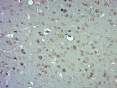
Paraformaldehyde-fixed, paraffin embedded (Mouse brain); Antigen retrieval by boiling in sodium citrate buffer (pH6.0) for 15min; Block endogenous peroxidase by 3% hydrogen peroxide for 20 minutes; Blocking buffer (normal goat serum) at 37°C for 30min; Antibody incubation with (Connexin 43) Polyclonal Antibody, Unconjugated (SL0651R) at 1:400 overnight at 4°C, followed by operating according to SP Kit(Rabbit) (sp-0023) instructionsand DAB staining.
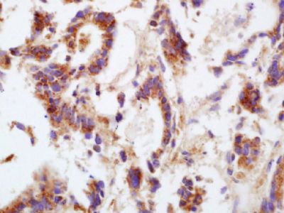
Paraformaldehyde-fixed, paraffin embedded (Human stomach cancer); Antigen retrieval by boiling in sodium citrate buffer (pH6.0) for 15min; Block endogenous peroxidase by 3% hydrogen peroxide for 20 minutes; Blocking buffer (normal goat serum) at 37°C for 30min; Antibody incubation with (Connexin 43) Polyclonal Antibody, Unconjugated (SL0651R) at 1:200 overnight at 4°C, followed by operating according to SP Kit(Rabbit) (sp-0023) instructions and DAB staining.
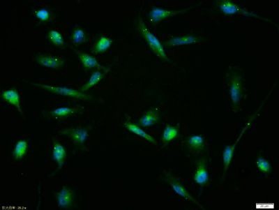
Tissue/cell:U-251 cell; 4% Paraformaldehyde-fixed; Triton X-100 at room temperature for 20 min; Blocking buffer (normal goat serum, SLC0005) at 37°C for 20 min; Antibody incubation with (Connexin 43) polyclonal Antibody, Unconjugated (SL0651R) 1:100, 90 minutes at 37°C; followed by a FITC conjugated Goat Anti-Rabbit IgG antibody at 37°C for 90 minutes, DAPI (blue, C02-04002) was used to stain the cell nuclei.
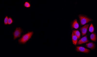
Tissue/cell: MCF7 cell; 4% Paraformaldehyde-fixed; Triton X-100 at room temperature for 20 min; Blocking buffer (normal goat serum, SLC0005) at 37°C for 20 min; Antibody incubation with (Connexin 43) polyclonal Antibody, Unconjugated (SL0651R) 1:100, 90 minutes at 37°C; followed by a FITC conjugated Goat Anti-Rabbit IgG antibody at 37°C for 90 minutes, DAPI (blue, C02-04002) was used to stain the cell nuclei.
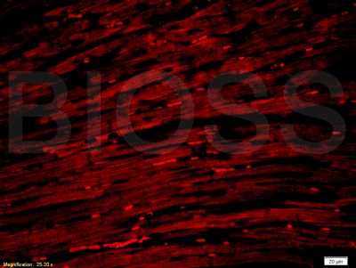
Tissue/cell: rat heart tissue;4% Paraformaldehyde-fixed and paraffin-embedded;
Antigen retrieval: citrate buffer ( 0.01M, pH 6.0 ), Boiling bathing for 15min; Blocking buffer (normal goat serum,SLC0005) at 37℃ for 20 min;
Incubation: Anti-Connexin 43 Polyclonal Antibody, Unconjugated(SL0651R) 1:200, overnight at 4°C; The secondary antibody was Goat Anti-Rabbit IgG, PE conjugated(SL0295G-PE)used at 1:200 dilution for 40 minutes at 37°C.
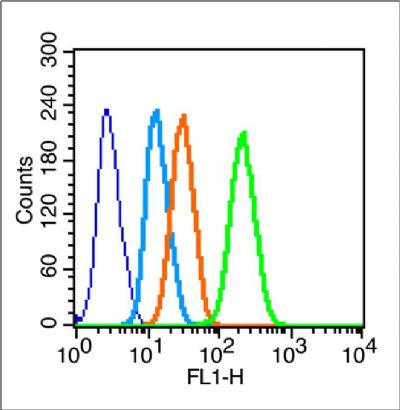
Blank control (blue line):Hela(blue).
Primary Antibody (green line): Rabbit Anti-Connexin 43 antibody(SL0651R)
Dilution: 1μg /10^6 cells;
Isotype Control Antibody (orange line): Rabbit IgG .
Secondary Antibody (white blue line): F(ab’)2 fragment goat anti-rabbit IgG-FITC.
Dilution: 1μg /test.
Protocol
The cells were fixed with 2% paraformaldehyde (10 min) , then permeabilized with 90% ice-cold methanol for 30 min on ice.Cells stained with Primary Antibody for 30 min at room temperature. The cells were then incubated in 1 X PBS/2%BSA/10% goat serum to block non-specific protein-protein interactions followed by the antibody for 15 min at room temperature. The secondary antibody used for 40 min at room temperature. Acquisition of 20,000 events was performed.
Product Feedback Wall
Message :
Your Email :
Copyright © 2007-2018 Sunlong Medical All Rights Reserved.