[IF=1.918] Jisheng Wang. et al. Effects of Diabetes Mellitus on Sperm Quality in the Db/Db Mouse Model and the Role of the FoxO1 Pathway. Med Sci Monitor. 2021; 27: e928232-1–e928232-12 WB ; Mouse.
[IF=1.487] Yuefeng Gaoet al. Vitamin E promotes ovine Sertoli cell proliferation by regulation of genes associated with cell division and the cell cycle. Anim Biotechnol
. 2020 Jul 2;1-9. IF ; sheep.
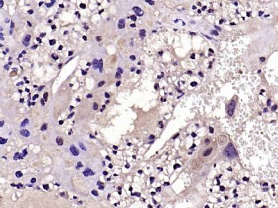
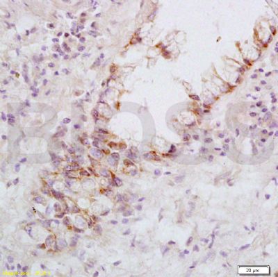
[IF=2.868] Jimbo H et al. Fas‐FasL interaction in cytotoxic T cell‐mediated vitiligo: The role of lesional expression of tumor necrosis factor‐α and interferon‐γ in Fas‐mediated melanocyte … Exp Dermatol. 2019 Nov 1. IHSLCP ; Human.
[IF=2.136] Liu S et al. PKCδ contributes to oxidative stress-induced apoptosis in porcine ovarian granulosa cells via activating JNK. Theriogenology. 2019 Jun;131:89-95. WB ; porcine.
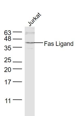
[IF=1.664] Wang Z et al. β-Bourbonene attenuates proliferation and induces apoptosis of prostate cancer cells.Oncol Lett. 2018 Oct;16(4):4519-4525. WB ; Human.
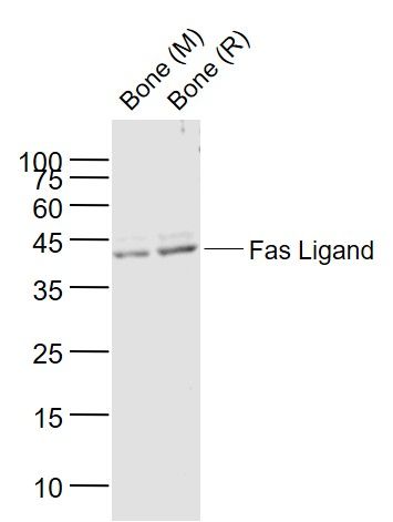
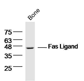
[IF=2.766] Zhang et al. Cytochrome P450 3A1 mediates 2,2',4,4'-tetrabromodiphenyl ether-induced reduction of spermatogenesis in adult rats. (2013) PLoS.One. 8:e66301 WB ; Rat.
[IF=5.23] Lv, Yanhong, et al. "Antiproliferative and Apoptosis-inducing Effect of exo-Protoporphyrin IX based Sonodynamic Therapy on Human Oral Squamous Cell Carcinoma." Scientific Reports 7 (2017): 40967. WB ; Mouse.
[IF=2.13] Shi, Yuqin, et al. "p, p′-DDE induces apoptosis of rat Sertoli cells via a FasL-dependent pathway." Journal of Biomedicine and Biotechnology (2009). Rat.
[IF=1.48] Wang, Shuhua, Qingqing Tian, and Fang An. "Growth inhibition and apoptotic effects of total flavonoids from Trollius chinensis on human breast cancer MCF-7 cells." Oncology Letters (2016). WB ; Human.
[IF=2.67] Wang, Yi, et al. "Lipopolysaccharide‐induced expression of FAS ligand in cultured immature boar Sertoli cells through the regulation of pro‐inflammatory cytokines and miR‐187." Molecular Reproduction and Development (2015). WB ;
[IF=3.53] Song, Xiufang, et al. "Polychlorinated biphenyl quinone metabolite promotes p53-dependent DNA damage checkpoint activation, S-phase cycle arrest and extrinsic apoptosis in human liver hepatocellular carcinoma HepG2 Cells."Chemical Research in Toxicology (2015). WB ; Human.
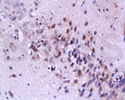
[IF=5.699] Janos L. Tanyi. et al. Personalized cancer vaccine strategy elicits polyfunctional T cells and demonstrates clinical benefits in ovarian cancer. Npj Vaccines. 2021 Mar;6(1):1-14 IHC ; Mouse.
[IF=1.77] Chen, Cui-Ying, et al. "Characterization of cytotoxicity-related gene expression in response to virulent Marek??s disease virus infection in the bursa of Fabricius."Research in veterinary science (2012).q WB ; Chicken.
[IF=2.87] Shi, Yuqin, et al. "β‐benzene hexachloride induces apoptosis of rat sertoli cells through generation of reactive oxygen species and activation of JNKs and FasL." Environmental toxicology 26.2 (2011): 124-135. WB ; Rat.
[IF=2.85] Qi, Suqin, et al. "BPA-induced apoptosis of rat Sertoli cells through Fas/FasL and JNKs/p38 MAPK pathways." Reproductive Toxicology 50 (2014): 108-116. WB ; Rat.