Home > Product > Antibody > Rabbit Anti-FLIP antibody
CASP8 and FADD-like apoptosis regulator subunit p43; CASP8 and FADD-like apoptosis regulator subunit p43; Flice-like Inhibitory protein; c FLIP; c FLIPL; c FLIPR; c FLIPS; c-FLIP; CASH; CASP8 and FADD like apoptosis regulator; CASP8 and FADD like apoptosi
Cat:
SL0119R
Species Reactivity:
Human,Mouse,Rat,(predicted: Dog,Pig,Cow,Rabbit,)
Immunogen:
KLH conjugated synthetic peptide derived from human CASP8 and FADD-like apoptosis regulator subunit p43:7-100/480
Format:
Liquid
Storage instructions:
Shipped at 4℃. Store at -20 °C for one year. Avoid repeated freeze/thaw cycles.
Concentration:
1mg/ml
Clonality:
Polyclonal
Isotype:
IgG
Applications:
WB=1:500-2000ELISA=1:5000-10000IHC-P=1:100-500IHC-F=1:100-500Flow-Cyt=1ug/TestIF=1:100-500(Paraffin sections need to do antigen repair)not yet tested in other applications.optimal dilutions/concentrations should be determined by the end user.
Host:
Rabbit
Product Overview:
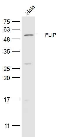
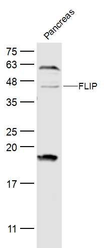
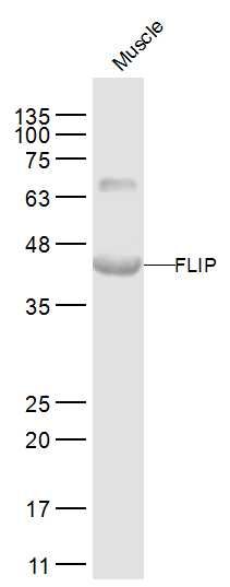
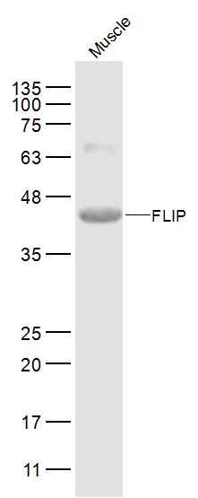
Sample: Hela(Human) Cell Lysate at 30 ugPrimary: Anti-FLIP (SL0119R) at 1/500 dilutionSecondary: IRDye800CW Goat Anti-Rabbit IgG at 1/20000 dilutionPredicted band size: 43/52 kDObserved band size: 52 kDSample: Pancreas (Mouse) Lysate at 40 ugPrimary: Anti-FLIP (SL0119R) at 1/500 dilutionSecondary: IRDye800CW Goat Anti-Rabbit IgG at 1/20000 dilutionPredicted band size: 43/52 kDObserved band size: 43/52 kDSample: Muscle(Mouse) Lysate at 40 ugPrimary:Anti-FLIP (SL0119R) at 1/2000 dilutionSecondary: IRDye800CW Goat Anti-Rabbit IgG at 1/20000 dilutionPredicted band size: 43/52 kDObserved band size: 43 kDSample: Muscle(Mouse) Lysate at 40 ugPrimary: Anti-FLIP (SL0119R) at 1/500 dilution Secondary: IRDye800CW Goat Anti-Rabbit IgG at 1/20000 dilutionPredicted band size: 43/52 kDObserved band size: 43 kDParaformaldehyde-fixed, paraffin embedded (rat liver tissue); Antigen retrieval by boiling in sodium citrate buffer (pH6.0) for 15min; Block endogenous peroxidase by 3% hydrogen peroxide for 20 minutes; Blocking buffer (normal goat serum) at 37°C for 30min; Antibody incubation with (FLIP) Polyclonal Antibody, Unconjugated (SL0199R) at 1:400 overnight at 4°C, followed by a conjugated secondary (sp-0023) for 20 minutes and DAB staining. Tissue/cell: rat brain tissue; 4% Paraformaldehyde-fixed and paraffin-embedded; Antigen retrieval: citrate buffer ( 0.01M, pH 6.0 ), Boiling bathing for 15min; Block endogenous peroxidase by 3% Hydrogen peroxide for 30min; Blocking buffer (normal goat serum,SLC0005) at 37℃ for 20 min; Incubation: Anti-FLIP/c FLIP Polyclonal Antibody, Unconjugated(SL0119R) 1:300, overnight at 4°C, followed by conjugation to the secondary antibody(SP-0023) and DAB(SLC0010) stainingBlank control: Hela. Primary Antibody (green line): Rabbit Anti-FLIP antibody (SL0119R) Dilution: 1ug/Test; Secondary Antibody : Goat anti-rabbit IgG-FITCDilution: 0.5ug/Test. ProtocolThe cells were fixed with 4% PFA (10min at room temperature)and then permeabilized with 90% ice-cold methanol for 20 min at -20℃.The cells were then incubated in 5%BSA to block non-specific protein-protein interactions for 30 min at room temperature .Cells stained with Primary Antibody for 30 min at room temperature. The secondary antibody used for 40 min at room temperature. Acquisition of 20,000 events was performed.Blank control (Black line): HUVEC (Black). Primary Antibody (green line): Rabbit Anti-FLIP antibody (SL0119R) Dilution: 1μg /10^6 cells; Isotype Control Antibody (orange line): Rabbit IgG . Secondary Antibody (white blue line): Goat anti-rabbit IgG-AF647Dilution: 1μg /test. ProtocolThe cells were fixed with 4% PFA (10min at room temperature)and then permeabilized with 0.1% PBST for 20 min at room temperature. The cells were then incubated in 5%BSA to block non-specific protein-protein interactions for 30 min at room temperature .Cells stained with Primary Antibody for 30 min at room temperature. The secondary antibody used for 40 min at room temperature. Acquisition of 20,000 events was performed.
More
Unit:
Price: $
Product PDFs
Datasheet:


The protein encoded by this gene is a regulator of apoptosis and is structurally similar to caspase-8. However, the encoded protein lacks caspase activity and appears to be itself cleaved into two peptides by caspase-8. Several transcript variants encoding different isoforms have been found for this gene, and partial evidence for several more variants exists. [provided by RefSeq, Feb 2011]

Function:
Apoptosis regulator protein which may function as a crucial link between cell survival and cell death pathways in mammalian cells. Acts as an inhibitor of TNFRSF6 mediated apoptosis. A proteolytic fragment (p43) is likely retained in the death-inducing signaling complex (DISC) thereby blocking further recruitment and processing of caspase-8 at the complex. Full length and shorter isoforms have been shown either to induce apoptosis or to reduce TNFRSF-triggered apoptosis. Lacks enzymatic (caspase) activity.

Subunit:
TNFRSF6 stimulation triggers recruitment to the death-inducing signaling complex (DISC) formed by TNFRSF6, FADD and caspase-8. A proteolytic fragment (p43) stays associated with the DISC. Also interacts with caspase-10, caspase-3, TRAF1, TRAF2 and Bcl-X(L) (in vitro). Interacts with HBV protein X.

Tissue Specificity:
Widely expressed. Higher expression in skeletal muscle, pancreas, heart, kidney, placenta, and peripheral blood leukocytes. Also detected in diverse cell lines. Isoform 8 is predominantly expressed in testis and skeletal muscle.

Post-translational modifications:
Proteolytically processed; probably by caspase-8. Processing likely occurs at the DISC and generates subunit p43 and p12.

Similarity:
Belongs to the peptidase C14A family.
Contains 2 DED (death effector) domains.

SWISS:
O15519

Gene ID:
8837

Database links:

Entrez Gene: 8837 Human

Entrez Gene: 12633 Mouse

Entrez Gene: 117279 Rat

Omim: 603599 Human

SwissProt: O15519 Human

SwissProt: O35732 Mouse

Unigene: 390736 Human

Unigene: 336848 Mouse


FLIP参与凋亡的调节。此抗体在长型和短型的FLIP异构体中均表达。短型FLIP包含2个死亡效应基因结构区,同源于FAS相关蛋白死亡效应基因结构区。长型FLIP包含1个附加的Caspase 样结构区,但是他缺少一个催化部位和在大多数Caspase 蛋白中形成底物结合束的残基。 Picture

Sample:
Hela(Human) Cell Lysate at 30 ug
Primary: Anti-FLIP (SL0119R) at 1/500 dilution
Secondary: IRDye800CW Goat Anti-Rabbit IgG at 1/20000 dilution
Predicted band size: 43/52 kD
Observed band size: 52 kD
Sample:
Pancreas (Mouse) Lysate at 40 ug
Primary: Anti-FLIP (SL0119R) at 1/500 dilution
Secondary: IRDye800CW Goat Anti-Rabbit IgG at 1/20000 dilution
Predicted band size: 43/52 kD
Observed band size: 43/52 kD
Sample:
Muscle(Mouse) Lysate at 40 ug
Primary:Anti-FLIP (SL0119R) at 1/2000 dilution
Secondary: IRDye800CW Goat Anti-Rabbit IgG at 1/20000 dilution
Predicted band size: 43/52 kD
Observed band size: 43 kD
Sample:
Muscle(Mouse) Lysate at 40 ug
Primary: Anti-FLIP (SL0119R) at 1/500 dilution
Secondary: IRDye800CW Goat Anti-Rabbit IgG at 1/20000 dilution
Predicted band size: 43/52 kD
Observed band size: 43 kD
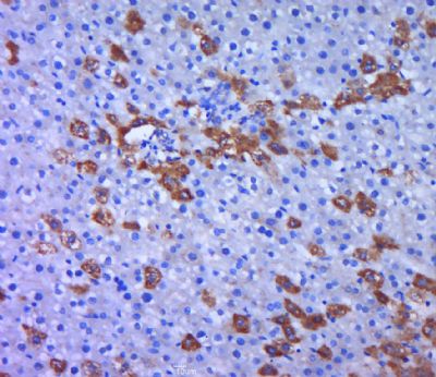
Paraformaldehyde-fixed, paraffin embedded (rat liver tissue); Antigen retrieval by boiling in sodium citrate buffer (pH6.0) for 15min; Block endogenous peroxidase by 3% hydrogen peroxide for 20 minutes; Blocking buffer (normal goat serum) at 37°C for 30min; Antibody incubation with (FLIP) Polyclonal Antibody, Unconjugated (SL0199R) at 1:400 overnight at 4°C, followed by a conjugated secondary (sp-0023) for 20 minutes and DAB staining.
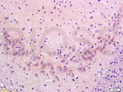
Tissue/cell: rat brain tissue; 4% Paraformaldehyde-fixed and paraffin-embedded;
Antigen retrieval: citrate buffer ( 0.01M, pH 6.0 ), Boiling bathing for 15min; Block endogenous peroxidase by 3% Hydrogen peroxide for 30min; Blocking buffer (normal goat serum,SLC0005) at 37℃ for 20 min;
Incubation: Anti-FLIP/c FLIP Polyclonal Antibody, Unconjugated(SL0119R) 1:300, overnight at 4°C, followed by conjugation to the secondary antibody(SP-0023) and DAB(SLC0010) staining
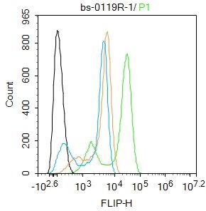
Blank control: Hela.
Primary Antibody (green line): Rabbit Anti-FLIP antibody (SL0119R)
Dilution: 1ug/Test;
Secondary Antibody : Goat anti-rabbit IgG-FITC
Dilution: 0.5ug/Test.
Protocol
The cells were fixed with 4% PFA (10min at room temperature)and then permeabilized with 90% ice-cold methanol for 20 min at -20℃.The cells were then incubated in 5%BSA to block non-specific protein-protein interactions for 30 min at room temperature .Cells stained with Primary Antibody for 30 min at room temperature. The secondary antibody used for 40 min at room temperature. Acquisition of 20,000 events was performed.
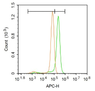
Blank control (Black line): HUVEC (Black).
Primary Antibody (green line): Rabbit Anti-FLIP antibody (SL0119R)
Dilution: 1μg /10^6 cells;
Isotype Control Antibody (orange line): Rabbit IgG .
Secondary Antibody (white blue line): Goat anti-rabbit IgG-AF647
Dilution: 1μg /test.
Protocol
The cells were fixed with 4% PFA (10min at room temperature)and then permeabilized with 0.1% PBST for 20 min at room temperature. The cells were then incubated in 5%BSA to block non-specific protein-protein interactions for 30 min at room temperature .Cells stained with Primary Antibody for 30 min at room temperature. The secondary antibody used for 40 min at room temperature. Acquisition of 20,000 events was performed.
Product Feedback Wall
Message :
Your Email :
Copyright © 2007-2018 Sunlong Medical All Rights Reserved.