Home > Product > Antibody > Rabbit Anti-GAP43 antibody
Growth Associated Protein-43; Neuromodulin; Axonal membrane protein GAP 43; B-50; F1; GAP 43; Growth Associated Protein 43; Nerve Growth Related Peptide; Neural phosphoprotein B 50; Neuromodulin; GAP-43; pp46; NEUM_HUMAN; Protein F1; QtrA-11580; QtrA-1307
Cat:
SL0154R
Species Reactivity:
Human,Mouse,Rat,(predicted: Chicken,Dog,)
Immunogen:
KLH conjugated synthetic peptide derived from human GAP43:9-100/238
Format:
Liquid
Storage instructions:
Shipped at 4℃. Store at -20 °C for one year. Avoid repeated freeze/thaw cycles.
Concentration:
1mg/ml
Clonality:
Polyclonal
Isotype:
IgG
Applications:
WB=1:500-2000ELISA=1:5000-10000IHC-P=1:100-500IHC-F=1:100-500IF=1:200-800(Paraffin sections need to do antigen repair)not yet tested in other applications.optimal dilutions/concentrations should be determined by the end user.
Host:
Rabbit
Product Overview:
Tissue/cell: rat brain tissue; 4% Paraformaldehyde-fixed and paraffin-embedded; Antigen retrieval: citrate buffer ( 0.01M, pH 6.0 ), Boiling bathing for 15min; Block endogenous peroxidase by 3% Hydrogen peroxide for 30min; Blocking buffer (normal goat serum,SLC0005) at 37℃ for 20 min; Incubation: Anti-GAP-43 Polyclonal Antibody, Unconjugated(SL0154R) 1:200, overnight at 4°C, followed by conjugation to the secondary antibody(SP-0023) and DAB(SLC0010) stainingParaformaldehyde-fixed, paraffin embedded (Human glioma); Antigen retrieval by boiling in sodium citrate buffer (pH6.0) for 15min; Block endogenous peroxidase by 3% hydrogen peroxide for 20 minutes; Blocking buffer (normal goat serum) at 37°C for 30min; Antibody incubation with (GAP43) Polyclonal Antibody, Unconjugated (SL0154R) at 1:400 overnight at 4°C, followed by a conjugated Goat Anti-Rabbit IgG antibody (SL0295G-FITC) for 90 minutes, and DAPI for nuclei staining.Paraformaldehyde-fixed, paraffin embedded (Mouse brain); Antigen retrieval by boiling in sodium citrate buffer (pH6.0) for 15min; Block endogenous peroxidase by 3% hydrogen peroxide for 20 minutes; Blocking buffer (normal goat serum) at 37°C for 30min; Antibody incubation with (GAP43) Polyclonal Antibody, Unconjugated (SL0154R) at 1:400 overnight at 4°C, followed by a conjugated Goat Anti-Rabbit IgG antibody (SL0295G-FITC) for 90 minutes, and DAPI for nuclei staining.Blank control (blue line): Hela cells (blue). Primary Antibody (green line): Rabbit Anti-GAP43 antibody (SL0154R) Dilution: 1μg /10^6 cells; Isotype Control Antibody (orange line): Rabbit IgG . Secondary Antibody (white blue line): Goat anti-rabbit IgG-PEDilution: 1μg /test. ProtocolThe cells were fixed with 70% methanol (Overnight at 4℃) and then permeabilized with 90% ice-cold methanol for 20 min at -20℃. Cells stained with Primary Antibody for 30 min at room temperature. The cells were then incubated in 1 X PBS/2%BSA/10% goat serum to block non-specific protein-protein interactions followed by the antibody for 15 min at room temperature. The secondary antibody used for 40 min at room temperature. Acquisition of 20,000 events was performed.
More
Unit:
Price: $
Product PDFs
Datasheet:


The protein encoded by this gene has been termed a 'growth' or 'plasticity' protein because it is expressed at high levels in neuronal growth cones during development and axonal regeneration. This protein is considered a crucial component of an effective regenerative response in the nervous system. Alternatively spliced transcript variants encoding distinct isoforms have been found for this gene. [provided by RefSeq, Jul 2008]

Function:
This protein is associated with nerve growth. It is a major component of the motile 'growth cones' that form the tips of elongating axons. Plays a role in axonal and dendritic filopodia induction.

Subunit:
Identified in a complex containing FGFR4, NCAM1, CDH2, PLCG1, FRS2, SRC, SHC1, GAP43 and CTTN. Binds calmodulin with a greater affinity in the absence of Ca(2+) than in its presence.

Subcellular Location:
Cell membrane; Peripheral membrane protein; Cytoplasmic side. Cell projection, growth cone membrane; Peripheral membrane protein; Cytoplasmic side. Cell junction, synapse. Cell projection, filopodium membrane; Peripheral membrane protein. Note=Cytoplasmic surface of growth cone and synaptic plasma membranes.

Post-translational modifications:
Phosphorylated at Ser-41 by PHK. Phosphorylation of this protein by a protein kinase C is specifically correlated with certain forms of synaptic plasticity.
Palmitoylation by ARF6 is essential for plasma membrane association and axonal and dendritic filopodia induction. Deacylated by LYPLA2.

Similarity:
Belongs to the neuromodulin family.
Contains 1 IQ domain.

SWISS:
P06837

Gene ID:
2596

Database links:

Entrez Gene: 2596 Human

Entrez Gene: 14432 Mouse

Entrez Gene: 29423 Rat

GenBank: NP_002036 Human

Omim: 162060 Human

SwissProt: P17677 Human

SwissProt: P06837 Mouse

SwissProt: P07936 Rat

Unigene: 134974 Human

Unigene: 1222 Mouse

Unigene: 10928 Rat


神经生物学相关蛋白(Neurobiology);神经标志物
GAP43 (Growth associated protein-43)又称作neuromodulin,是一个轴突膜蛋白,是一种神经特异性的蛋白质,参与神经细胞外生长及突触发育形成和神经细胞再生。在神经元发育和再生过程中以高水平表达。能调解轴突延伸作用,改变细胞形态。作为细胞内信号,可大大增强与G蛋白偶联的受体转运作用。
神经生长相关蛋白-GAP-43和神经细胞黏附因子(neural cell adhesion molecule,NCAM)与突触可塑性密切相关.GAP-43是一种神经细胞膜上的特异性磷蛋白,在神经发育和再生过程中呈现高表达,被作为突触生长的标志物,有称脊髓生长相关蛋白
GAP-43与CaM结合,参与G蛋白相互作用,神经递质的释放,作用于胞吞/胞吐过程,通过小囊溶合或诱导生长锥和突触前末端的胞吞促进膜扩展,与海马长时程增强密切相关.
神经细胞黏附因子是细胞表面糖蛋白大家族的成员之一,促进轴突生长,对长时记忆的保持有重要影响,同时,GAP-43对其具调节作用,广泛而深入地研究GAP-43意义深远。 Picture

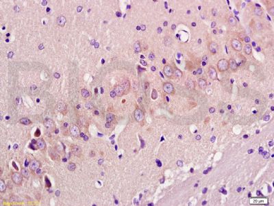
Tissue/cell: rat brain tissue; 4% Paraformaldehyde-fixed and paraffin-embedded;
Antigen retrieval: citrate buffer ( 0.01M, pH 6.0 ), Boiling bathing for 15min; Block endogenous peroxidase by 3% Hydrogen peroxide for 30min; Blocking buffer (normal goat serum,SLC0005) at 37℃ for 20 min;
Incubation: Anti-GAP-43 Polyclonal Antibody, Unconjugated(SL0154R) 1:200, overnight at 4°C, followed by conjugation to the secondary antibody(SP-0023) and DAB(SLC0010) staining
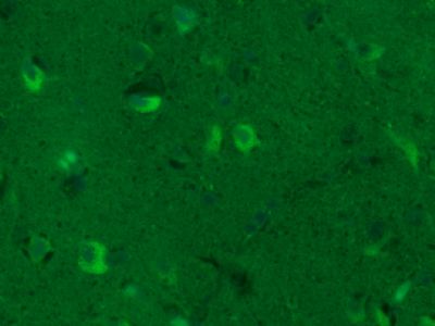
Paraformaldehyde-fixed, paraffin embedded (Human glioma); Antigen retrieval by boiling in sodium citrate buffer (pH6.0) for 15min; Block endogenous peroxidase by 3% hydrogen peroxide for 20 minutes; Blocking buffer (normal goat serum) at 37°C for 30min; Antibody incubation with (GAP43) Polyclonal Antibody, Unconjugated (SL0154R) at 1:400 overnight at 4°C, followed by a conjugated Goat Anti-Rabbit IgG antibody (SL0295G-FITC) for 90 minutes, and DAPI for nuclei staining.
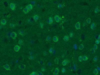
Paraformaldehyde-fixed, paraffin embedded (Mouse brain); Antigen retrieval by boiling in sodium citrate buffer (pH6.0) for 15min; Block endogenous peroxidase by 3% hydrogen peroxide for 20 minutes; Blocking buffer (normal goat serum) at 37°C for 30min; Antibody incubation with (GAP43) Polyclonal Antibody, Unconjugated (SL0154R) at 1:400 overnight at 4°C, followed by a conjugated Goat Anti-Rabbit IgG antibody (SL0295G-FITC) for 90 minutes, and DAPI for nuclei staining.
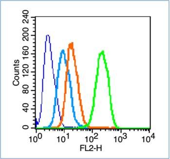
Blank control (blue line): Hela cells (blue).
Primary Antibody (green line): Rabbit Anti-GAP43 antibody (SL0154R)
Dilution: 1μg /10^6 cells;
Isotype Control Antibody (orange line): Rabbit IgG .
Secondary Antibody (white blue line): Goat anti-rabbit IgG-PE
Dilution: 1μg /test.
Protocol
The cells were fixed with 70% methanol (Overnight at 4℃) and then permeabilized with 90% ice-cold methanol for 20 min at -20℃. Cells stained with Primary Antibody for 30 min at room temperature. The cells were then incubated in 1 X PBS/2%BSA/10% goat serum to block non-specific protein-protein interactions followed by the antibody for 15 min at room temperature. The secondary antibody used for 40 min at room temperature. Acquisition of 20,000 events was performed.
Product Feedback Wall
Message :
Your Email :
Copyright © 2007-2018 Sunlong Medical All Rights Reserved.