Home > Product > Antibody > Rabbit Anti-HSP60 antibody
HSP 60; HSP-60; 60 kDa chaperonin;60 kDa heat shock protein mitochondrial; 60kDa; cb863; Chaperonin 60; Chaperonin;Chaperonin, 60-KD; CPN 60; CPN60; fa04a05; fb22d10;fi27b05; GROEL; GroEL Homolog; GroEL, E. coli, homolog of; Heat shock 60kD protein 1 (cha
Cat:
SL0191R
Species Reactivity:
Human,Mouse,Rat,(predicted: Dog,Cow,Horse,Rabbit,)
Immunogen:
KLH conjugated synthetic peptide derived from human HSP60:501-573/573
Format:
Liquid
Storage instructions:
Shipped at 4℃. Store at -20 °C for one year. Avoid repeated freeze/thaw cycles.
Concentration:
1mg/ml
Clonality:
Polyclonal
Isotype:
IgG
Applications:
WB=1:500-2000ELISA=1:5000-10000IHC-P=1:100-500IHC-F=1:100-500ICC=1:100IF=1:100-500(Paraffin sections need to do antigen repair)not yet tested in other applications.optimal dilutions/concentrations should be determined by the end user.
Host:
Rabbit
Product Overview:
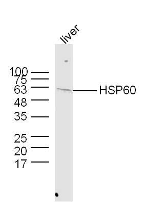
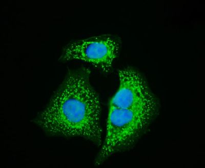
Sample: Kidney (Mouse) Lysate at 30 ugPrimary: Anti- HSP60 (SL0191R) at 1/300 dilutionSecondary: IRDye800CW Goat Anti-Rabbit IgG at 1/20000 dilutionPredicted band size: 58 kDObserved band size: 58 kDSample: Heart (Mouse) Lysate at 30 ugPrimary: Anti-HSP60 (SL0191R) at 1/300 dilutionSecondary: IRDye800CW Goat Anti-Rabbit IgG at 1/20000 dilutionPredicted band size: 58 kDObserved band size: 58 kDSample: Lane 1: Liver (Mouse) Lysate at 40 ugLane 2: Adrenal gland (Mouse) Lysate at 40 ugLane 3: NIH/3T3 (Mouse) Cell Lysate at 30 ugLane 4: Adrenal gland (Rat) Lysate at 40 ugLane 5: Jurkat (Human) Cell Lysate at 30 ugLane 6: MDA-MB-231 (Human) Cell Lysate at 30 ugLane 7: HepG2 (Human) Cell Lysate at 30 ugLane 8: Hela (Human) Cell Lysate at 30 ugLane 9: K562 (Human) Cell Lysate at 30 ugPrimary: Anti-HSP60 (SL0191R) at 1/1000 dilutionSecondary: IRDye800CW Goat Anti-Rabbit IgG at 1/20000 dilutionPredicted band size: 62 kDObserved band size: 62 kDSample: Hela(Human) Cell Lysate at 30 ugPrimary: Anti- HSP60 (SL0191R) at 1/1000 dilutionSecondary: IRDye800CW Goat Anti-Rabbit IgG at 1/20000 dilutionPredicted band size: 58 kDObserved band size: 58 kDSample: Liver (Mouse) Lysate at 40 ugPrimary: Anti-HSP60 (SL0191R) at 1/300 dilutionSecondary: IRDye800CW Goat Anti-Rabbit IgG at 1/20000 dilutionPredicted band size: 58 kDObserved band size: 58 kDParaformaldehyde-fixed, paraffin embedded (rat stomach); Antigen retrieval by boiling in sodium citrate buffer (pH6.0) for 15min; Block endogenous peroxidase by 3% hydrogen peroxide for 20 minutes; Blocking buffer (normal goat serum) at 37°C for 30min; Antibody incubation with (HSP60) Polyclonal Antibody, Unconjugated (SL0191R) at 1:500 overnight at 4°C, followed by a conjugated secondary (sp-0023) for 20 minutes and DAB staining.Tissue/cell: rat brain tissue; 4% Paraformaldehyde-fixed and paraffin-embedded; Antigen retrieval: citrate buffer ( 0.01M, pH 6.0 ), Boiling bathing for 15min; Block endogenous peroxidase by 3% Hydrogen peroxide for 30min; Blocking buffer (normal goat serum,SLC0005) at 37∩ for 20 min; Incubation: Anti-HSP60 Polyclonal Antibody, Unconjugated(SL0191R) 1:200, overnight at 4∑C, followed by conjugation to the secondary antibody(SP-0023) and DAB(SLC0010) stainingTissue/cell: human lung carcinoma; 4% Paraformaldehyde-fixed and paraffin-embedded; Antigen retrieval: citrate buffer ( 0.01M, pH 6.0 ), Boiling bathing for 15min; Block endogenous peroxidase by 3% Hydrogen peroxide for 30min; Blocking buffer (normal goat serum,SLC0005) at 37∩ for 20 min; Incubation: Anti-HSP60 Polyclonal Antibody, Unconjugated(SL0191R) 1:200, overnight at 4∑C, followed by conjugation to the secondary antibody(SP-0023) and DAB(SLC0010) stainingHepG2 cell; 4% Paraformaldehyde-fixed; Triton X-100 at room temperature for 20 min; Blocking buffer (normal goat serum, SLC0005) at 37°C for 20 min; Antibody incubation with (HSP60) polyclonal Antibody, Unconjugated (SL0191R) 1:100, 90 minutes at 37°C; followed by a conjugated Goat Anti-Rabbit IgG antibody at 37°C for 90 minutes, DAPI (blue, C02-04002) was used to stain the cell nuclei.
More
Unit:
Price: $
Product PDFs
Datasheet:


This gene encodes a member of the chaperonin family. The encoded mitochondrial protein may function as a signaling molecule in the innate immune system. This protein is essential for the folding and assembly of newly imported proteins in the mitochondria. This gene is adjacent to a related family member and the region between the 2 genes functions as a bidirectional promoter. Two pseudogenes, both located on chromosome 8, have been associated with this gene. Two transcript variants encoding the same protein have been identified for this gene. Mutations associated with this gene cause autosomal recessive spastic paraplegia 13.

Function:
Implicated in mitochondrial protein import and macromolecular assembly. May facilitate the correct folding of imported proteins. May also prevent misfolding and promote the refolding and proper assembly of unfolded polypeptides generated under stress conditions in the mitochondrial matrix.

Subunit:
Interacts with HRAS (By similarity). Interacts with HBV protein X and HTLSLV1 protein p40tax. Interacts with ATAD3A.

Subcellular Location:
Mitochondrion matrix.

DISEASE:
Spastic paraplegia autosomal dominant 13 (SPG13) [MIM:605280]: A form of spastic paraplegia, a neurodegenerative disorder characterized by a slow, gradual, progressive weakness and spasticity of the lower limbs. Rate of progression and the severity of symptoms are quite variable. Initial symptoms may include difficulty with balance, weakness and stiffness in the legs, muscle spasms, and dragging the toes when walking. In some forms of the disorder, bladder symptoms (such as incontinence) may appear, or the weakness and stiffness may spread to other parts of the body. Note=The disease is caused by mutations affecting the gene represented in this entry.
Leukodystrophy, hypomyelinating, 4 (HLD4) [MIM:612233]: A severe autosomal recessive hypomyelinating leukodystrophy. Clinically characterized by infantile-onset rotary nystagmus, progressive spastic paraplegia, neurologic regression, motor impairment, profound mental retardation. Death usually occurs within the first two decades of life. Note=The disease is caused by mutations affecting the gene represented in this entry.

Similarity:
Belongs to the chaperonin (HSP60) family.

SWISS:
P10809

Gene ID:
3329

Database links:

Entrez Gene: 3329 Human

Entrez Gene: 15510 Mouse

Entrez Gene: 63868 Rat

Omim: 118190 Human

SwissProt: P86206 Hamster

SwissProt: P10809 Human

SwissProt: P63038 Mouse

SwissProt: P63039 Rat

Unigene: 595053 Human

Unigene: 727543 Human

Unigene: 1777 Mouse

Unigene: 102058 Rat


信号传导(Signaling Intermediates)
HSP60是一类进化上高度保守的蛋白质家族.生理状态时协助多肽或蛋白质的正确转位、折叠和装配,起"分子伴侣"的作用;在应激状态下,HSP60过表达或异位表达,作为一种自身抗原被免疫系统识别,诱发机体的保护性免疫应答,也可作为一种信号分子,在信号转导中发挥作用.
该蛋白与肿瘤的发生、增殖及分化有关,主要用于舌鳞癌、结肠癌、胰腺癌和乳腺癌的研究,近年来研究证实HSP60在自身免疫性疾病、传染病、动脉粥样硬化及慢性感染的发病中均发挥重要的作用。
HSP-60热休克蛋白-蛋白质折叠过程的引导因子

分子伴侣是一种引导蛋白质正确折叠的蛋白质。当蛋白质折叠时,它们能保护蛋白质分子免受其它蛋白质的干扰。很多分子伴侣属于热休克蛋白(例如HSP-60),它们在细胞受热时大量合成。热激可导致蛋白质稳定性降低,增加错误折叠的几率,因此在受到热刺激时,细胞中的蛋白质需要更多热休克蛋白的帮助。
HSP-60是一种典型的分子伴侣,它可以为正在折叠的蛋白质提供一个附着环境,从而起到保护折叠过程的作用。 Picture

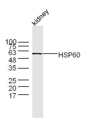
Sample: Kidney (Mouse) Lysate at 30 ug
Primary: Anti- HSP60 (SL0191R) at 1/300 dilution
Secondary: IRDye800CW Goat Anti-Rabbit IgG at 1/20000 dilution
Predicted band size: 58 kD
Observed band size: 58 kD
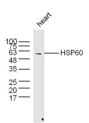
Sample: Heart (Mouse) Lysate at 30 ug
Primary: Anti-HSP60 (SL0191R) at 1/300 dilution
Secondary: IRDye800CW Goat Anti-Rabbit IgG at 1/20000 dilution
Predicted band size: 58 kD
Observed band size: 58 kD
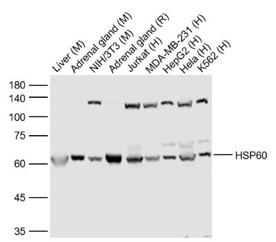
Sample:
Lane 1: Liver (Mouse) Lysate at 40 ug
Lane 2: Adrenal gland (Mouse) Lysate at 40 ug
Lane 3: NIH/3T3 (Mouse) Cell Lysate at 30 ug
Lane 4: Adrenal gland (Rat) Lysate at 40 ug
Lane 5: Jurkat (Human) Cell Lysate at 30 ug
Lane 6: MDA-MB-231 (Human) Cell Lysate at 30 ug
Lane 7: HepG2 (Human) Cell Lysate at 30 ug
Lane 8: Hela (Human) Cell Lysate at 30 ug
Lane 9: K562 (Human) Cell Lysate at 30 ug
Primary: Anti-HSP60 (SL0191R) at 1/1000 dilution
Secondary: IRDye800CW Goat Anti-Rabbit IgG at 1/20000 dilution
Predicted band size: 62 kD
Observed band size: 62 kD
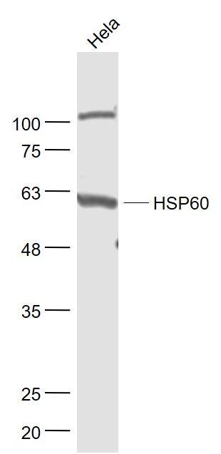
Sample:
Hela(Human) Cell Lysate at 30 ug
Primary: Anti- HSP60 (SL0191R) at 1/1000 dilution
Secondary: IRDye800CW Goat Anti-Rabbit IgG at 1/20000 dilution
Predicted band size: 58 kD
Observed band size: 58 kD
Sample:
Liver (Mouse) Lysate at 40 ug
Primary: Anti-HSP60 (SL0191R) at 1/300 dilution
Secondary: IRDye800CW Goat Anti-Rabbit IgG at 1/20000 dilution
Predicted band size: 58 kD
Observed band size: 58 kD
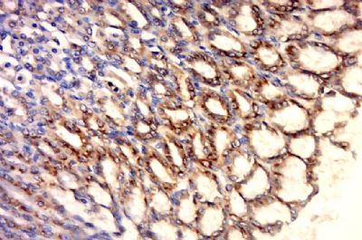
Paraformaldehyde-fixed, paraffin embedded (rat stomach); Antigen retrieval by boiling in sodium citrate buffer (pH6.0) for 15min; Block endogenous peroxidase by 3% hydrogen peroxide for 20 minutes; Blocking buffer (normal goat serum) at 37°C for 30min; Antibody incubation with (HSP60) Polyclonal Antibody, Unconjugated (SL0191R) at 1:500 overnight at 4°C, followed by a conjugated secondary (sp-0023) for 20 minutes and DAB staining.
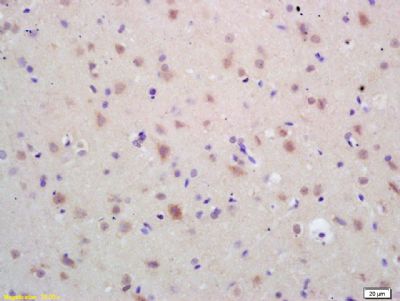
Tissue/cell: rat brain tissue; 4% Paraformaldehyde-fixed and paraffin-embedded;
Antigen retrieval: citrate buffer ( 0.01M, pH 6.0 ), Boiling bathing for 15min; Block endogenous peroxidase by 3% Hydrogen peroxide for 30min; Blocking buffer (normal goat serum,SLC0005) at 37∩ for 20 min;
Incubation: Anti-HSP60 Polyclonal Antibody, Unconjugated(SL0191R) 1:200, overnight at 4∑C, followed by conjugation to the secondary antibody(SP-0023) and DAB(SLC0010) staining
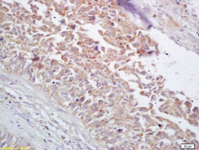
Tissue/cell: human lung carcinoma; 4% Paraformaldehyde-fixed and paraffin-embedded;
Antigen retrieval: citrate buffer ( 0.01M, pH 6.0 ), Boiling bathing for 15min; Block endogenous peroxidase by 3% Hydrogen peroxide for 30min; Blocking buffer (normal goat serum,SLC0005) at 37∩ for 20 min;
Incubation: Anti-HSP60 Polyclonal Antibody, Unconjugated(SL0191R) 1:200, overnight at 4∑C, followed by conjugation to the secondary antibody(SP-0023) and DAB(SLC0010) staining
HepG2 cell; 4% Paraformaldehyde-fixed; Triton X-100 at room temperature for 20 min; Blocking buffer (normal goat serum, SLC0005) at 37°C for 20 min; Antibody incubation with (HSP60) polyclonal Antibody, Unconjugated (SL0191R) 1:100, 90 minutes at 37°C; followed by a conjugated Goat Anti-Rabbit IgG antibody at 37°C for 90 minutes, DAPI (blue, C02-04002) was used to stain the cell nuclei.
Product Feedback Wall
Message :
Your Email :
Copyright © 2007-2018 Sunlong Medical All Rights Reserved.