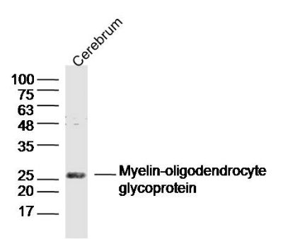
Sample:
Cerebrum (Mouse) Lysate at 40 ug
Primary: Anti- Myelin-oligodendrocyte glycoprotein (SL0426R) at 1/300 dilution
Secondary: IRDye800CW Goat Anti-Rabbit IgG at 1/20000 dilution
Predicted band size: 24 kD
Observed band size: 26 kD
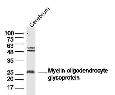
Sample:
Cerebrum (Rat) Lysate at 40 ug
Primary: Anti- Myelin-oligodendrocyte glycoprotein (SL0426R) at 1/300 dilution
Secondary: IRDye800CW Goat Anti-Rabbit IgG at 1/20000 dilution
Predicted band size: 24 kD
Observed band size: 26 kD
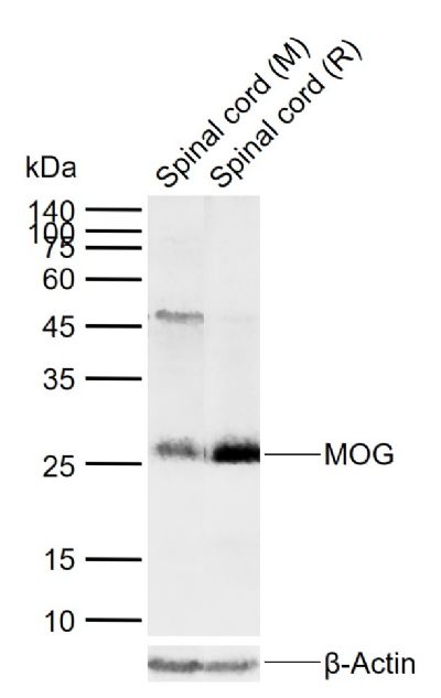
Sample:
Lane 1: Mouse Spinal cord tissue lysates
Lane 2: Rat Spinal cord tissue lysates
Primary: Anti-MOG (SL0426R) at 1/1000 dilution
Secondary: IRDye800CW Goat Anti-Rabbit IgG at 1/20000 dilution
Predicted band size: 24 kDa
Observed band size: 26 kDa
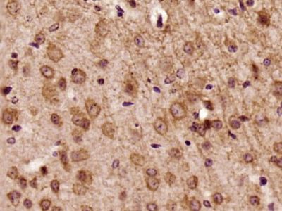
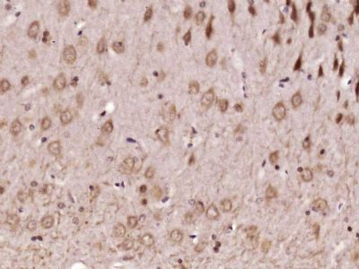
Paraformaldehyde-fixed, paraffin embedded (Mouse brain); Antigen retrieval by boiling in sodium citrate buffer (pH6.0) for 15min; Block endogenous peroxidase by 3% hydrogen peroxide for 20 minutes; Blocking buffer (normal goat serum) at 37°C for 30min; Antibody incubation with (MOG) Polyclonal Antibody, Unconjugated (SL0426R) at 1:500 overnight at 4°C, followed by a conjugated secondary (sp-0023) for 20 minutes and DAB staining.
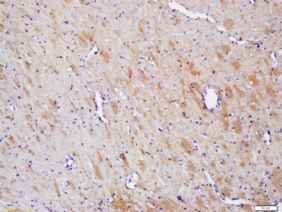
Paraformaldehyde-fixed, paraffin embedded (Rat brain); Antigen retrieval by boiling in sodium citrate buffer (pH6.0) for 15min; Block endogenous peroxidase by 3% hydrogen peroxide for 20 minutes; Blocking buffer (normal goat serum) at 37°C for 30min; Antibody incubation with (MOG) Polyclonal Antibody, Unconjugated (SL0426R) at 1:500 overnight at 4°C, followed by a conjugated secondary (sp-0023) for 20 minutes and DAB staining.
Tissue/cell: rat brain tissue; 4% Paraformaldehyde-fixed and paraffin-embedded;
Antigen retrieval: citrate buffer ( 0.01M, pH 6.0 ), Boiling bathing for 15min; Block endogenous peroxidase by 3% Hydrogen peroxide for 30min; Blocking buffer (normal goat serum,SLC0005) at 37← for 20 min;
Incubation: Anti- MOG Polyclonal Antibody, Unconjugated(SL0426R) 1:200, overnight at 4⒉C, followed by conjugation to the secondary antibody(SP-0023) and DAB(SLC0010) staining
|