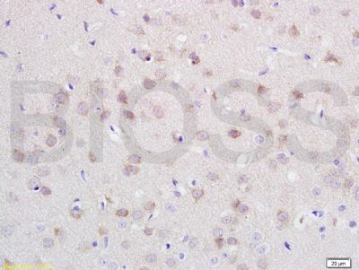
[IF=5.923] Nanami Nakamura. et al. Possible Action of Olaparib for Preventing Invasion of Oral Squamous Cell Carcinoma In Vitro and In Vivo. Int J Mol Sci. 2022 Jan;23(5):2527 WB ; Mouse.
[IF=4.101] Haixiang Ding. et al. Ailanthone suppresses the activity of human colorectal cancer cells through the STAT3 signaling pathway. Int J Mol Med. 2022 Feb;49(2):1-11 WB ; Human.
[IF=4.784] Zheng Wu. et al. FOXD3 suppresses epithelial–mesenchymal transition through direct transcriptional promotion of SMAD7 in esophageal squamous cell carcinoma. 2021 Sep 22 WB ; human.
[IF=6.023] Ling Xie. et al. Suppression of GOLM1 by EGCG through HGF/HGFR/AKT/GSK-3β/β-catenin/c-Myc signaling pathway inhibits cell migration of MDA-MB-231. Food Chem Toxicol. 2021 Nov;157:112574 WB ; human.
[IF=9.933] Xingyi Xu. et al. A Honeycomb-Like Bismuth/Manganese Oxide Nanoparticle with Mutual Reinforcement of Internal and External Response for Triple-Negative Breast Cancer Targeted Therapy. 2021 Jul 23 WB ; Human.
[IF=4.432] Botao Pan. et al. Vorinostat targets UBE2C to reverse epithelial-mesenchymal transition and control cervical cancer growth through the ubiquitination pathway. Eur J Pharmacol. 2021 Oct;908:174399 WB ; Human.
[IF=7.076] Mengmeng Niu. et al. Noncanonical TGF-β signaling leads to FBXO3-mediated degradation of ΔNp63α promoting breast cancer metastasis and poor clinical prognosis. Plos Biol. 2021 Feb;19(2):e3001113 WB ; Human.
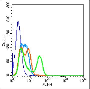
[IF=3.391] Jasmine Ercoliet al. KRIT1 as a possible new player in melanoma aggressiveness. Arch Biochem Biophys
. 2020 Sep 30;691:108483. WB ; Human.
[IF=5.572] Liu H et al. Anti-tubulin agent vinorelbine inhibits metastasis of cancer cells by regulating epithelial-mesenchymal transition. Eur J Med Chem. 2020 Aug 15;200:112332. WB ; Human.
[IF=2.058] Ma T et al. MiR-940 inhibits migration and invasion of tongue squamous cell carcinoma via regulatingCXCR2/NF-κB system-mediated epithelial–mesenchymal transition. Naunyn Schmiedebergs Arch Pharmacol. 2019 Jun 18. WB&IF ; Human.
[IF=2.705] Zhou S et al. Helicobacter pylori infection promotes epithelial-to-mesenchymal transition of gastric cells by upregulating LAPTM4B. Biochem Biophys Res Commun. 2019 Jun 30;514(3):893-900. IHSLCP,ICF&WB ; Human.
[IF=4.357] Fei et al. The number of polyploid giant cancer cells and epithelial-mesenchymal transition-related proteins are associated with invasion and metastasis in human breast cancer. (2015) J.Exp.Clin.Cancer.Res. 34:158 WB ; Human.
[IF=2.766] Ning et al. Vascular endothelial growth factor receptor-1 activation promotes migration and invasion of breast cancer cells through epithelial-mesenchymal transition. (2013) PLoS.One. 8:e65217 IHSLCP ; Human.
[IF=3.26] Park, Honghyun, Doyun Kim, and Kuen Yong Lee. "Interaction‐tailored cell aggregates in alginate hydrogels for enhanced chondrogenic differentiation."Journal of Biomedical Materials Research Part A (2016). IHSLCP ; Mouse.
[IF=4.36] Yuhua Li. et al. Bolbostemma paniculatum (Maxim.) Franquet extract suppresses the development of colorectal cancer through downregulation of PI3K/Akt pathway. J Ethnopharmacol. 2022 Apr;287:114937 WB ; Human.
[IF=6.691] Zhao, Boyuan. et al. Suspension state and shear stress enhance breast tumor cells EMT through YAP by microRNA-29b. 2021 Oct 07 WB ; Human.
[IF=4.101] Yu Guo. et al. RepSox effectively promotes the induced differentiation of sheep fibroblasts into adipocytes via the inhibition of the TGF‑β1/Smad pathway. Int J Mol Med. 2021 Aug;48(2):1-13 WB ; Sheep.
[IF=4.147] Francesca Salamanna. et al. Development and characterization of a novel human 3D model of bone metastasis from breast carcinoma in vitro cultured. Bone. 2021 Feb;143:115773 IHC ; Human.
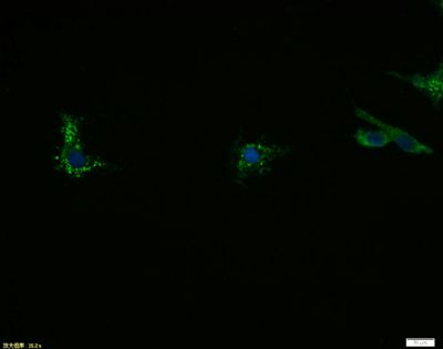
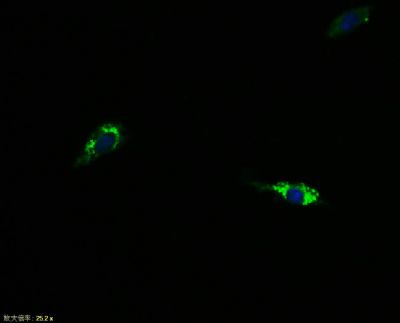
[IF=7.727] Xue Wang. et al. Engineered liposomes targeting the gut–CNS Axis for comprehensive therapy of spinal cord injury. J Control Release. 2021 Mar;331:390 IF ; Rat.
[IF=3.98] Liu, Wen-bin, et al. "Aberrant methylation accounts for cell adhesion-related gene silencing during 3-methylcholanthrene and diethylnitrosamine induced multistep rat lung carcinogenesis associated with overexpression of DNA methyltransferases 1 and 3a." Toxicology and applied pharmacology 251.1 (2011): 70-78. IHSLCP ; Rat.
[IF=8.456] Zhang D et al. Cell Membrane-Coated Porphyrin Metal-Organic Frameworks for Cancer Cell Targeting and O2-Evolving Photodynamic Therapy. ACS Appl Mater Interfaces. 2019 Oct 30;11(43):39594-39602. WB ; Human.
[IF=3.405] Wu Y et al. MFAP5 promotes basal-like breast cancer progression by activating the EMT program.Cell Biosci. 2019 Mar 7;9:24. WB ; Human.
[IF=6.217] Zhang J, et al. LGR5, a novel functional glioma stem cell marker, promotes EMT by activating the Wnt/β-catenin pathway and predicts poor survival of glioma patients.(2018) J. Exp. Clin. Cancer Res. Sep 12;37(1):225. IHC ; Human.
[IF=4.12] Wang et al. Kukoamine A inhibits human glioblastoma cell growth and migration through apoptosis induction and epithelial-mesenchymal transition attenuation. (2016) Sci.Rep. 6:36543 WB ; Human.
[IF=2.2] Yang, Wei, et al. "lncRNA H19 is involved in TGF-β1-induced epithelial to mesenchymal transition in bovine epithelial cells through PI3K/AKT Signaling Pathway." PeerJ 5 (2017): e3950. WB ; Bovine.