Home > Product > Antibody > Rabbit Anti-NSE antibody
Gamma-enolase; Gamma enolase; Neural enolase; Neuron specific enolase; neurone-specific enolase; 2 phospho D glycerate hydrolyase; Eno 2; Eno2; ENO2; ENOG; Enolase 2; Enolase 2 gamma neuronal; Enolase2; Neuron specific enolase; Neuron specific gamma enola
Cat:
SL1027R
Species Reactivity:
Human,Mouse,Rat,Dog,(predicted: Chicken,Cow,)
Immunogen:
KLH conjugated synthetic peptide derived from human NSE:201-300/434
Format:
Liquid
Storage instructions:
Shipped at 4℃. Store at -20 °C for one year. Avoid repeated freeze/thaw cycles.
Concentration:
1mg/ml
Clonality:
Polyclonal
Isotype:
IgG
Applications:
WB=1:500-2000ELISA=1:5000-10000IHC-P=1:100-500IHC-F=1:100-500Flow-Cyt=1μg/Test IF=1:100-500(Paraffin sections need to do antigen repair)not yet tested in other applications.optimal dilutions/concentrations should be determined by the end user.
Host:
Rabbit
Product Overview:
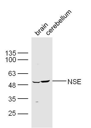
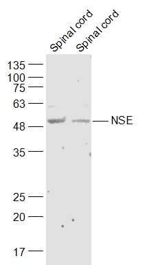
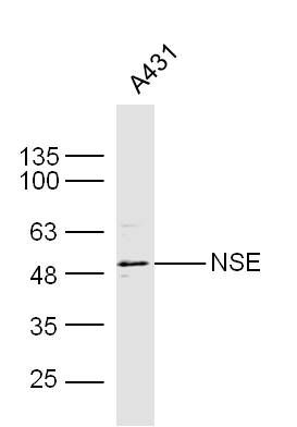
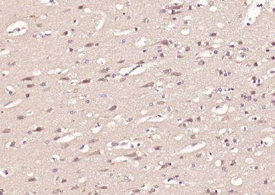
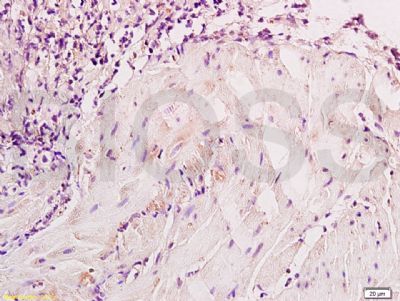
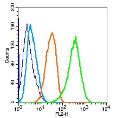
Sample: Brain (Mouse) Lysate at 30 ugCerebellum (Mouse) Lysate at 30 ugPrimary: Anti-NSE (SL1027R) at 1/300 dilutionSecondary: IRDye800CW Goat Anti-Rabbit IgG at 1/20000 dilutionPredicted band size: 48 kDObserved band size: 49kDSample: Spinal cord (Mouse) Lysate at 40 ugSpinal cord (Rat) Lysate at 40 ugPrimary: Anti-NSE (SL1027R) at 1/300 dilutionSecondary: IRDye800CW Goat Anti-Rabbit IgG at 1/20000 dilutionPredicted band size: 48 kDObserved band size: 48 kDSample: A431 Cell Lysate at 40 ugPrimary: Anti- NSE (SL1027R) at 1/300 dilutionSecondary: IRDye800CW Goat Anti-Rabbit IgG at 1/20000 dilutionPredicted band size: 48 kDObserved band size: 48 kDParaformaldehyde-fixed, paraffin embedded (human brain); Antigen retrieval by boiling in sodium citrate buffer (pH6.0) for 15min; Block endogenous peroxidase by 3% hydrogen peroxide for 20 minutes; Blocking buffer (normal goat serum) at 37°C for 30min; Antibody incubation with (NSE) Polyclonal Antibody, Unconjugated (SL1027R) at 1:200 overnight at 4°C, followed by operating according to SP Kit(Rabbit) (sp-0023) instructionsand DAB staining.Paraformaldehyde-fixed, paraffin embedded (rat brain); Antigen retrieval by boiling in sodium citrate buffer (pH6.0) for 15min; Block endogenous peroxidase by 3% hydrogen peroxide for 20 minutes; Blocking buffer (normal goat serum) at 37°C for 30min; Antibody incubation with (NSE) Polyclonal Antibody, Unconjugated (SL1027R) at 1:200 overnight at 4°C, followed by operating according to SP Kit(Rabbit) (sp-0023) instructionsand DAB staining.Paraformaldehyde-fixed, paraffin embedded (mouse brain); Antigen retrieval by boiling in sodium citrate buffer (pH6.0) for 15min; Block endogenous peroxidase by 3% hydrogen peroxide for 20 minutes; Blocking buffer (normal goat serum) at 37°C for 30min; Antibody incubation with (NSE) Polyclonal Antibody, Unconjugated (SL1027R) at 1:200 overnight at 4°C, followed by operating according to SP Kit(Rabbit) (sp-0023) instructionsand DAB staining. Paraformaldehyde-fixed, paraffin embedded (Rat brain); Antigen retrieval by boiling in sodium citrate buffer (pH6.0) for 15min; Block endogenous peroxidase by 3% hydrogen peroxide for 20 minutes; Blocking buffer (normal goat serum) at 37°C for 30min; Antibody incubation with (NSE) Polyclonal Antibody, Unconjugated (SL1027R) at 1:500 overnight at 4°C, followed by a conjugated secondary (sp-0023) for 20 minutes and DAB staining. Tissue/cell: Neuroblastoma cells; Block endogenous peroxidase by 3% Hydrogen peroxide for 30min; Blocking buffer (normal goat serum,SLC0005) at 37℃ for 20 min; Incubation: Anti-NSE/ENO2/γ Enolase Polyclonal Antibody, Unconjugated(SL1027R) 1:200, overnight at 4°C, followed by conjugation to the secondary antibody(SP-0023) and DAB(SLC0010) stainingTissue/cell: dog bladder tissue; 4% Paraformaldehyde-fixed and paraffin-embedded; Antigen retrieval: citrate buffer ( 0.01M, pH 6.0 ), Boiling bathing for 15min; Block endogenous peroxidase by 3% Hydrogen peroxide for 30min; Blocking buffer (normal goat serum,SLC0005) at 37℃ for 20 min; Incubation: Anti-NSE/ENO2/γ Enolase Polyclonal Antibody, Unconjugated(SL1027R) 1:800, overnight at 4°C, followed by conjugation to the secondary antibody(SP-0023) and DAB(SLC0010) stainingBlank control: U-87MG(blue). Primary Antibody:Rabbit Anti-NSE antibody(SL1027R), Dilution: 1μg in 100 μL 1X PBS containing 0.5% BSA; Isotype Control Antibody: Rabbit IgG(orange) ,used under the same conditions ); Secondary Antibody: Goat anti-rabbit IgG-PE(white blue), Dilution: 1:200 in 1 X PBS containing 0.5% BSA.ProtocolThe cells were fixed with 2% paraformaldehyde (10 min) , then permeabilized with 90% ice-cold methanol for 30 min on ice. Primary antibody (SL1027R,1μg /1x10^6 cells) were incubated for 30 min on the ice, followed by 1 X PBS containing 0.5% BSA + 1 0% goat serum (15 min) to block non-specific protein-protein interactions. Then the Goat Anti-rabbit IgG/PE antibody was added into the blocking buffer mentioned above to react with the primary antibody at 1/200 dilution for 30 min on ice. Acquisition of 20,000 events was performed.
More
Unit:
Price: $
Product PDFs
Datasheet:


This gene encodes one of the three enolase isoenzymes found in mammals. This isoenzyme, a homodimer, is found in mature neurons and cells of neuronal origin. A switch from alpha enolase to gamma enolase occurs in neural tissue during development in rats and primates. [provided by RefSeq, Jul 2008].

Function:
Has neurotrophic and neuroprotective properties on a broad spectrum of central nervous system (CNS) neurons. Binds, in a calcium-dependent manner, to cultured neocortical neurons and promotes cell survival.

Subunit:
Mammalian enolase is composed of 3 isozyme subunits, alpha, beta and gamma, which can form homodimers or heterodimers which are cell-type and development-specific.

Subcellular Location:
Cytoplasm. Cell membrane. Can translocate to the plasma membrane in either the homodimeric (alpha/alpha) or heterodimeric (alpha/gamma) form.

Tissue Specificity:
The alpha/alpha homodimer is expressed in embryo and in most adult tissues. The alpha/beta heterodimer and the beta/beta homodimer are found in striated muscle, and the alpha/gamma heterodimer and the gamma/gamma homodimer in neurons.

Similarity:
Belongs to the enolase family.

SWISS:
P09104

Gene ID:
2026

Database links:

Entrez Gene: 2026 Human

Entrez Gene: 2767 Mouse

Entrez Gene: 24334 Rat

Omim: 13172 Human

SwissProt: P09104 Human

SwissProt: P17183 Mouse

SwissProt: P07323 Rat

Unigene: 511915 Human


神经元特异性烯醇化酶(NSE)存在于神经元细胞和神经内分泌组织中,起源于神经内分泌细胞的肿瘤可产生过量的NSE。NSE也是小细胞肺癌的检测指标,70%左右的小细胞肺癌患者血中NSE升高,而其他组织型肺癌NSE升高的患者仅为10%~20%。 Picture

Sample:
Brain (Mouse) Lysate at 30 ug
Cerebellum (Mouse) Lysate at 30 ug
Primary: Anti-NSE (SL1027R) at 1/300 dilution
Secondary: IRDye800CW Goat Anti-Rabbit IgG at 1/20000 dilution
Predicted band size: 48 kD
Observed band size: 49kD
Sample:
Spinal cord (Mouse) Lysate at 40 ug
Spinal cord (Rat) Lysate at 40 ug
Primary: Anti-NSE (SL1027R) at 1/300 dilution
Secondary: IRDye800CW Goat Anti-Rabbit IgG at 1/20000 dilution
Predicted band size: 48 kD
Observed band size: 48 kD
Sample: A431 Cell Lysate at 40 ug
Primary: Anti- NSE (SL1027R) at 1/300 dilution
Secondary: IRDye800CW Goat Anti-Rabbit IgG at 1/20000 dilution
Predicted band size: 48 kD
Observed band size: 48 kD
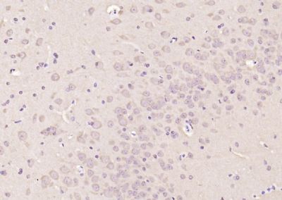
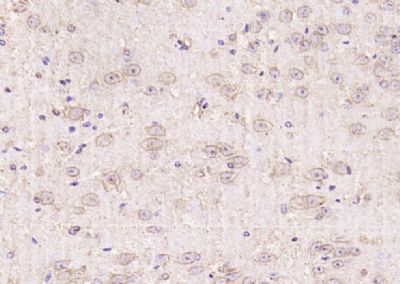
Paraformaldehyde-fixed, paraffin embedded (human brain); Antigen retrieval by boiling in sodium citrate buffer (pH6.0) for 15min; Block endogenous peroxidase by 3% hydrogen peroxide for 20 minutes; Blocking buffer (normal goat serum) at 37°C for 30min; Antibody incubation with (NSE) Polyclonal Antibody, Unconjugated (SL1027R) at 1:200 overnight at 4°C, followed by operating according to SP Kit(Rabbit) (sp-0023) instructionsand DAB staining.
Paraformaldehyde-fixed, paraffin embedded (rat brain); Antigen retrieval by boiling in sodium citrate buffer (pH6.0) for 15min; Block endogenous peroxidase by 3% hydrogen peroxide for 20 minutes; Blocking buffer (normal goat serum) at 37°C for 30min; Antibody incubation with (NSE) Polyclonal Antibody, Unconjugated (SL1027R) at 1:200 overnight at 4°C, followed by operating according to SP Kit(Rabbit) (sp-0023) instructionsand DAB staining.
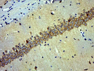
Paraformaldehyde-fixed, paraffin embedded (mouse brain); Antigen retrieval by boiling in sodium citrate buffer (pH6.0) for 15min; Block endogenous peroxidase by 3% hydrogen peroxide for 20 minutes; Blocking buffer (normal goat serum) at 37°C for 30min; Antibody incubation with (NSE) Polyclonal Antibody, Unconjugated (SL1027R) at 1:200 overnight at 4°C, followed by operating according to SP Kit(Rabbit) (sp-0023) instructionsand DAB staining.
Paraformaldehyde-fixed, paraffin embedded (Rat brain); Antigen retrieval by boiling in sodium citrate buffer (pH6.0) for 15min; Block endogenous peroxidase by 3% hydrogen peroxide for 20 minutes; Blocking buffer (normal goat serum) at 37°C for 30min; Antibody incubation with (NSE) Polyclonal Antibody, Unconjugated (SL1027R) at 1:500 overnight at 4°C, followed by a conjugated secondary (sp-0023) for 20 minutes and DAB staining.
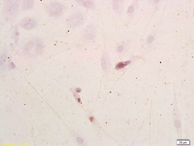
Tissue/cell: Neuroblastoma cells;
Block endogenous peroxidase by 3% Hydrogen peroxide for 30min; Blocking buffer (normal goat serum,SLC0005) at 37℃ for 20 min;
Incubation: Anti-NSE/ENO2/γ Enolase Polyclonal Antibody, Unconjugated(SL1027R) 1:200, overnight at 4°C, followed by conjugation to the secondary antibody(SP-0023) and DAB(SLC0010) staining
Tissue/cell: dog bladder tissue; 4% Paraformaldehyde-fixed and paraffin-embedded;
Antigen retrieval: citrate buffer ( 0.01M, pH 6.0 ), Boiling bathing for 15min; Block endogenous peroxidase by 3% Hydrogen peroxide for 30min; Blocking buffer (normal goat serum,SLC0005) at 37℃ for 20 min;
Incubation: Anti-NSE/ENO2/γ Enolase Polyclonal Antibody, Unconjugated(SL1027R) 1:800, overnight at 4°C, followed by conjugation to the secondary antibody(SP-0023) and DAB(SLC0010) staining
Blank control: U-87MG(blue).
Primary Antibody:Rabbit Anti-NSE antibody(SL1027R), Dilution: 1μg in 100 μL 1X PBS containing 0.5% BSA;
Isotype Control Antibody: Rabbit IgG(orange) ,used under the same conditions );
Secondary Antibody: Goat anti-rabbit IgG-PE(white blue), Dilution: 1:200 in 1 X PBS containing 0.5% BSA.
Protocol
The cells were fixed with 2% paraformaldehyde (10 min) , then permeabilized with 90% ice-cold methanol for 30 min on ice. Primary antibody (SL1027R,1μg /1x10^6 cells) were incubated for 30 min on the ice, followed by 1 X PBS containing 0.5% BSA + 1 0% goat serum (15 min) to block non-specific protein-protein interactions. Then the Goat Anti-rabbit IgG/PE antibody was added into the blocking buffer mentioned above to react with the primary antibody at 1/200 dilution for 30 min on ice. Acquisition of 20,000 events was performed.
Product Feedback Wall
Message :
Your Email :
Copyright © 2007-2018 Sunlong Medical All Rights Reserved.