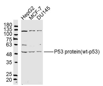
Sample:
HepG2 Cell Lysate at 40 ug
Mcf-7 Cell Lysate at 40 ug
DU145 Cell Lysate at 40 ug
Primary: Anti- P53 protein(wt-p53)(SL0033R) at 1/300 dilution
Secondary: IRDye800CW Goat Anti-Rabbit IgG at 1/20000 dilution
Predicted band size: 43 kD
Observed band size: 50 kD
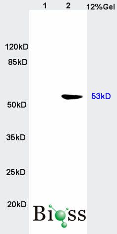
Sample:
Brain(Rat) lysate at 30ug;
Colon carcinoma(Human) lysate at30 ug;
Primary: Anti-wt-p53 (SL0033R) at 1:200 dilution;
Secondary: HRP conjugated Goat-Anti-Rabbit IgG(SL0295G) at 1: 3000 dilution;
Predicted band size : 43kD
Observed band size : 53kD
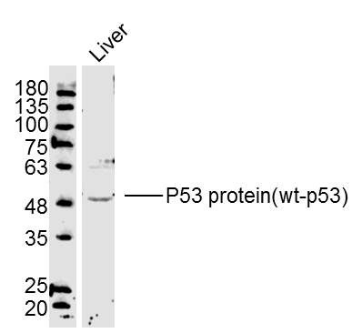
Sample:Liver(Mouse) Lysate at 40 ug
Primary: Anti-P53 protein(wt-p53)(SL0033R) at 1/300 dilution
Secondary: IRDye800CW Goat Anti-Rabbit IgG at 1/20000 dilution
Predicted band size: 43 kD
Observed band size: 50 kD
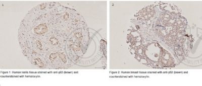
Independently Validated Antibody, image provided by Science Direct, badge number 029660. Formalin-fixed and paraffin embedded human testis and breast tissue stained with Rabbit Anti-P53 protein(wt-p53) Polyclonal Antibody at 1:250 at room temperature overnight. Both positive and negative controls stained.
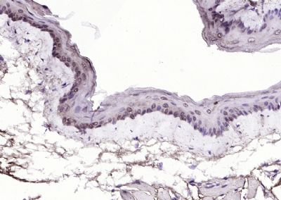
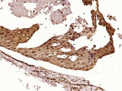
Paraformaldehyde-fixed, paraffin embedded (mouse esophageal); Antigen retrieval by boiling in sodium citrate buffer (pH6.0) for 15min; Block endogenous peroxidase by 3% hydrogen peroxide for 20 minutes; Blocking buffer (normal goat serum) at 37°C for 30min; Antibody incubation with (P53 protein(wt-p53)) Polyclonal Antibody, Unconjugated (SL0033R) at 1:200 overnight at 4°C, followed by operating according to SP Kit(Rabbit) (sp-0023) instructions and DAB staining.
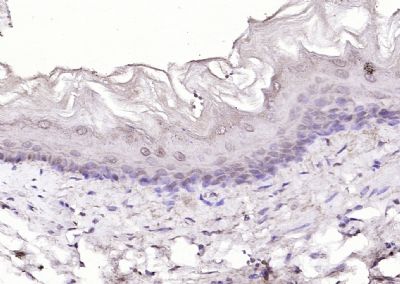
Paraformaldehyde-fixed, paraffin embedded (Rat esophageal); Antigen retrieval by boiling in sodium citrate buffer (pH6.0) for 15min; Block endogenous peroxidase by 3% hydrogen peroxide for 20 minutes; Blocking buffer (normal goat serum) at 37°C for 30min; Antibody incubation with (P53 protein(wt-p53)) Polyclonal Antibody, Unconjugated (SL0033R) at 1:200 overnight at 4°C, followed by operating according to SP Kit(Rabbit) (sp-0023) instructions and DAB staining.
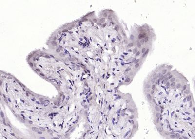
Paraformaldehyde-fixed, paraffin embedded (Rat bladder); Antigen retrieval by boiling in sodium citrate buffer (pH6.0) for 15min; Block endogenous peroxidase by 3% hydrogen peroxide for 20 minutes; Blocking buffer (normal goat serum) at 37°C for 30min; Antibody incubation with (P53 protein(wt-p53)) Polyclonal Antibody, Unconjugated (SL0033R) at 1:200 overnight at 4°C, followed by operating according to SP Kit(Rabbit) (sp-0023) instructions and DAB staining.
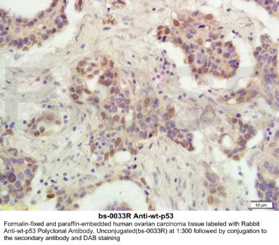
Paraformaldehyde-fixed, paraffin embedded (Human breast cancer); Antigen retrieval by boiling in sodium citrate buffer (pH6.0) for 15min; Block endogenous peroxidase by 3% hydrogen peroxide for 20 minutes; Blocking buffer (normal goat serum) at 37°C for 30min; Antibody incubation with (P53 protein(wt-p53)) Polyclonal Antibody, Unconjugated (SL0033R) at 1:400 overnight at 4°C, followed by operating according to SP Kit(Rabbit) (sp-0023) instructions and DAB staining.
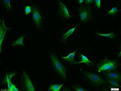
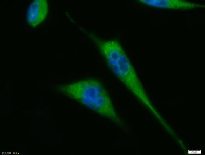
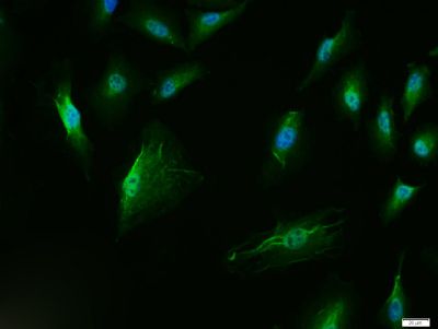
Tissue/cell: A549 cell; 4% Paraformaldehyde-fixed; Triton X-100 at room temperature for 20 min; Blocking buffer (normal goat serum, SLC0005) at 37°C for 20 min; Antibody incubation with (P53 protein(wt-p53)) polyclonal Antibody, Unconjugated (SL0033R) 1:100, 90 minutes at 37°C; followed by a FITC conjugated Goat Anti-Rabbit IgG antibody at 37°C for 90 minutes, DAPI (blue, C02-04002) was used to stain the cell nuclei.
Tissue/cell: A431 cell; 4% Paraformaldehyde-fixed; Triton X-100 at room temperature for 20 min; Blocking buffer (normal goat serum, SLC0005) at 37°C for 20 min; Antibody incubation with (P53 protein(wt-p53)) polyclonal Antibody, Unconjugated (SL0033R) 1:100, 90 minutes at 37°C; followed by a FITC conjugated Goat Anti-Rabbit IgG antibody at 37°C for 90 minutes, DAPI (blue, C02-04002) was used to stain the cell nuclei.
Tissue/cell: A549 cell; 4% Paraformaldehyde-fixed; Triton X-100 at room temperature for 20 min; Blocking buffer (normal goat serum, SLC0005) at 37°C for 20 min; Antibody incubation with (P53 protein(wt-p53)) polyclonal Antibody, Unconjugated (SL0033R) 1:100, 90 minutes at 37°C; followed by a FITC conjugated Goat Anti-Rabbit IgG antibody at 37°C for 90 minutes, DAPI (blue, C02-04002) was used to stain the cell nuclei.
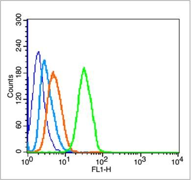
Blank control (blue line):HeLa(blue).
Primary Antibody (green line): Rabbit Anti-P53 protein(wt-p53) antibody(SL0033R),
Dilution: 1μg /10^6 cells;
Isotype Control Antibody (orange line): Rabbit IgG .
Secondary Antibody (white blue line): Goat anti-rabbit IgG-FITC
Dilution: 1μg /test.
Protocol
The cells were fixed with 70% ethanol (Overnight at 4℃) and then permeabilized with 90% ice-cold methanol for 30 min on ice. Cells stained with Primary Antibody for 30 min at room temperature. The cells were then incubated in 1 X PBS/2%BSA/10% goat serum to block non-specific protein-protein interactions followed by the antibody for 15 min at room temperature. The secondary antibody used for 40 min at room temperature. Acquisition of 20,000 events was performed.
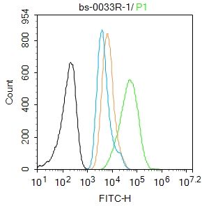
Blank control:A549.
Primary Antibody (green line): Rabbit Anti-P53 protein(wt-p53) antibody (SL0033R)
Dilution: 1μg /10^6 cells;
Isotype Control Antibody (orange line): Rabbit IgG .
Secondary Antibody : Goat anti-rabbit IgG-AF488
Dilution: 1μg /test.
Protocol
The cells were fixed with 4% PFA (10min at room temperature)and then permeabilized with 90% ice-cold methanol for 20 min at -20℃. The cells were then incubated in 5%BSA to block non-specific protein-protein interactions for 30 min at room temperature .Cells stained with Primary Antibody for 30 min at room temperature. The secondary antibody used for 40 min at room temperature. Acquisition of 20,000 events was performed.
|