[IF=9.933] Laijun Liu. et al. Hierarchical Nanostructured Electrospun Membrane with Periosteum-Mimic Microenvironment for Enhanced Bone Regeneration. 2021 Aug 05 IF ; Rat.
[IF=7.328] Dongdong Yao. et al. Matrix stiffness regulates bone repair by modulating 12-lipoxygenase-mediated early inflammation. Mat Sci Eng SLCMater. 2021 Sep;128:112359 IF ; Rat.
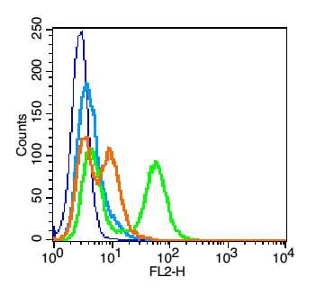
[IF=6.331] Shuying Hu. et al. Superparamagnetic core–shell electrospun scaffolds with sustained release of IONPs facilitating in vitro and in vivo bone regeneration. J Mater Chem B. 2021 Aug;: IF ; Rat.
[IF=5.157] Ge Liu. et al. Marine bacterial exopolysaccharide EPS11 inhibits migration and invasion of liver cancer cells by directly targeting collagen I. J Biol Chem. 2021 Aug;:101133 WB ; Human.
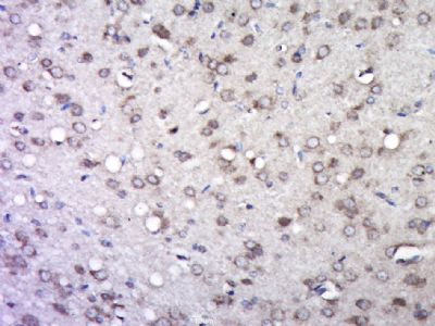
[IF=8.352] Chanjuan Dong. et al. Graphene-based conductive fibrous scaffold boosts sciatic nerve regeneration and functional recovery upon electrical stimulation. Appl Mater Today. 2020 Dec;21:100870 IHC ; Rat.
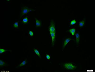
[IF=1.41] Lu T et al. In vitro culture and biological properties of broiler adipose‑derived stem cells.Exp Ther Med. 2018 Sep;16(3):2399-487. ICF&FITC ; broiler.
[IF=3.55] Fu, Min, et al. "Inositol Hexaphosphate and Inositol Inhibit Colorectal Cancer Metastasis to the Liver in BALB/c Mice." Nutrients 8.5 (2016): 286. IHSLCP ; Mouse.
[IF=3.27] Oralova, V., et al. "Osteogenic Potential of the Transcription Factor c-MYB." Calcified Tissue International: 1-12. WB ; Mouse.
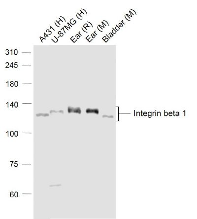
[IF=2.09] Qin, Jie, et al. "Concentrated growth factor promotes Schwann cell migration partly through the integrin β1-mediated activation of the focal adhesion kinase pathway." International Journal of Molecular Medicine. WB ; Rat.
[IF=1.89] Deng, Wei, et al. "Bone marrow mesenchymal stromal cells with CD47 high expression via the signal transducer and activators of transcription signaling pathway preventing myocardial fibrosis." International Journal of Clinical and Experimental Pathology 8.9 (2015): 10555-10564. other ; Rat.
[IF=3.58] Lei, Mingxing, et al. "Modulating hair follicle size with Wnt10b/DKK1 during hair regeneration." Experimental Dermatology (2014). IHSLCP ; Mouse.
[IF=5.168] Wang et al. Loss of calponin h1 confers anoikis resistance and tumor progression in the development of high-grade serous carcinoma originating from the fallopian tube epithelium. (2017) Oncotarget. 8:61133-61145 WB ; Human.
[IF=5.923] Xingyu Shen. et al. Extracellular Calcium Ion Concentration Regulates Chondrocyte Elastic Modulus and Adhesion Behavior. Int J Mol Sci. 2021 Jan;22(18):10034 IF ; huamn.
[IF=1.329] Ziwei Luo. et al. miR‐203a‐3p promotes loureirin A‐induced hair follicle stem cells differentiation by targeting Smad1. Anat Rec. 2021 Mar;304(3):531-540 IF ; Rat.
[IF=2.388] Heng Chen. et al. Podosome formation in the murine palatal mucosae: Its proteolytic role in rete peg formation. Ann Anat. 2021 May;235:151703 IF ; Mouse.
[IF=2.24] Chen, Xiaodong, et al. "The effect of porcine ADM to improve the burn wound healing." Int J Clin Exp Pathol 6.11 (2013): 2280-2291. WB ; Rat.
[IF=2.53] Ji, Kaiyuan, et al. "MicroRNA 143‐5p regulates alpaca melanocyte migration, proliferation, and melanogenesis." Experimental dermatology (2017). WB ; Other Species.
[IF=2.02] Zhao, Xue, et al. "Knockdown of Snail inhibits epithelial-mesenchymal transition of human laryngeal squamous cell carcinoma Hep-2 cells through VDR signaling pathway." Biochemistry and Cell Biology ja (2017). WB ; Human.