Home > Product > Antibody > Rabbit Anti-CHEK2 antibody
CHK2_HUMAN; Serine/threonine-protein kinase Chk2; EC:2.7.11.1; Serine/threonine-protein kinase Chk2; CDS1; CHK2; LFS2; RAD53; hCds1; HuCds1; PP1425; CHK2 checkpoint homolog; Cds1 homolog (Hucds1; hCds1); Checkpoint kinase 2;
Cat:
SL1391R
Species Reactivity:
Human,(predicted: Mouse,Rat,Dog,Cow,Horse,Rabbit,)
Immunogen:
KLH conjugated synthetic peptide derived from human CHEK2:101-250/586
Format:
Liquid
Storage instructions:
Shipped at 4℃. Store at -20 °C for one year. Avoid repeated freeze/thaw cycles.
Concentration:
1mg/ml
Clonality:
Polyclonal
Isotype:
IgG
Applications:
WB=1:500-2000ELISA=1:5000-10000IHC-P=1:100-500IHC-F=1:100-500Flow-Cyt=1ug/TesIF=1:100-500(Paraffin sections need to do antigen repair)not yet tested in other applications.optimal dilutions/concentrations should be determined by the end user.
Host:
Rabbit
Product Overview:
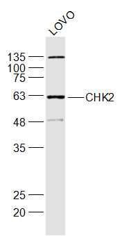
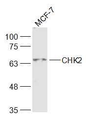
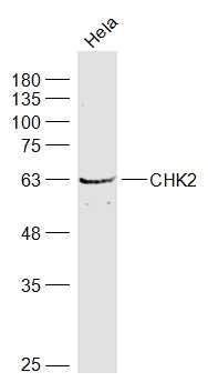
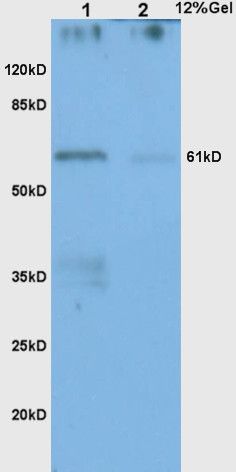
Sample: LOVO(Human) Cell Lysate at 30 ugPrimary: Anti-CHK2 (SL1391R) at 1/1000 dilutionSecondary: IRDye800CW Goat Anti-Rabbit IgG at 1/20000 dilutionPredicted band size: 65 kDObserved band size: 63 kDSample: MCF-7(Human) Cell Lysate at 30 ugPrimary: Anti-CHK2 (SL1391R) at 1/1000 dilutionSecondary: IRDye800CW Goat Anti-Rabbit IgG at 1/20000 dilutionPredicted band size: 65 kDObserved band size: 65 kDSample: Hela(Human) Cell Lysate at 30 ugPrimary: Anti-CHK2 (SL1391R) at 1/1000 dilutionSecondary: IRDye800CW Goat Anti-Rabbit IgG at 1/20000 dilutionPredicted band size: 65 kDObserved band size: 65 kDSample: Lane1: Colon carcinoma (Human) Lysate at 30 ugLane2: Gastric carcinoma(Human) Lysate at 30 ugPrimary: Anti-CHK2 (SL1391R) at 1:200 dilution; Secondary: HRP conjugated Goat Anti-Rabbit IgG(SL0295G-HRP) at 1: 3000 dilution; Predicted band size : 65kDObserved band size : 61kD Paraformaldehyde-fixed, paraffin embedded (Human breast carcinoma); Antigen retrieval by boiling in sodium citrate buffer (pH6.0) for 15min; Block endogenous peroxidase by 3% hydrogen peroxide for 20 minutes; Blocking buffer (normal goat serum) at 37°C for 30min; Antibody incubation with (CHK2) Polyclonal Antibody, Unconjugated (SL1391R) at 1:400 overnight at 4°C, followed by operating according to SP Kit(Rabbit) (sp-0023) instructionsand DAB staining.Tissue/cell: human gastric carcinoma; 4% Paraformaldehyde-fixed and paraffin-embedded; Antigen retrieval: citrate buffer ( 0.01M, pH 6.0 ), Boiling bathing for 15min; Block endogenous peroxidase by 3% Hydrogen peroxide for 30min; Blocking buffer (normal goat serum,SLC0005) at 37℃ for 20 min; Incubation: Anti-CHK2 Polyclonal Antibody, Unconjugated(SL1391R) 1:200, overnight at 4°C, followed by conjugation to the secondary antibody(SP-0023) and DAB(SLC0010) stainingBlank control: A431. Primary Antibody (green line): Rabbit Anti-CHK2 antibody (SL1391R) Dilution: 1ug/Test; Secondary Antibody : Goat anti-rabbit IgG-FITCDilution: 0.5ug/Test. ProtocolThe cells were fixed with 4% PFA (10min at room temperature)and then permeabilized with 90% ice-cold methanol for 20 min at -20℃.The cells were then incubated in 5%BSA to block non-specific protein-protein interactions for 30 min at room temperature .Cells stained with Primary Antibody for 30 min at room temperature. The secondary antibody used for 40 min at room temperature. Acquisition of 20,000 events was performed.Blank control: A431. Primary Antibody (green line): Rabbit Anti-CHK2 antibody (SL1391R) Dilution: 1μg /10^6 cells; Isotype Control Antibody (orange line): Rabbit IgG . Secondary Antibody : Goat anti-rabbit IgG-AF647Dilution: 1μg /test. ProtocolThe cells were fixed with 4% PFA (10min at room temperature)and then permeabilized with 90% ice-cold methanol for 20 min at-20℃. The cells were then incubated in 5%BSA to block non-specific protein-protein interactions for 30 min at at room temperature .Cells stained with Primary Antibody for 30 min at room temperature. The secondary antibody used for 40 min at room temperature. Acquisition of 20,000 events was performed.
More
Unit:
Price: $
Product PDFs
Datasheet:


In response to DNA damage and replication blocks, cell cycle progression is halted through the control of critical cell cycle regulators. The protein encoded by this gene is a cell cycle checkpoint regulator and putative tumor suppressor. It contains a forkhead-associated protein interaction domain essential for activation in response to DNA damage and is rapidly phosphorylated in response to replication blocks and DNA damage. When activated, the encoded protein is known to inhibit CDC25C phosphatase, preventing entry into mitosis, and has been shown to stabilize the tumor suppressor protein p53, leading to cell cycle arrest in G1. In addition, this protein interacts with and phosphorylates BRCA1, allowing BRCA1 to restore survival after DNA damage. Mutations in this gene have been linked with Li-Fraumeni syndrome, a highly penetrant familial cancer phenotype usually associated with inherited mutations in TP53. Also, mutations in this gene are thought to confer a predisposition to sarcomas, breast cancer, and brain tumors. This nuclear protein is a member of the CDS1 subfamily of serine/threonine protein kinases. Several transcript variants encoding different isoforms have been found for this gene. [provided by RefSeq, Apr 2012]

Function:
Serine/threonine-protein kinase which is required for checkpoint-mediated cell cycle arrest, activation of DNA repair and apoptosis in response to the presence of DNA double-strand breaks. May also negatively regulate cell cycle progression during unperturbed cell cycles. Following activation, phosphorylates numerous effectors preferentially at the consensus sequence [L-X-R-X-X-S/T]. Regulates cell cycle checkpoint arrest through phosphorylation of CDC25A, CDC25B and CDC25C, inhibiting their activity. Inhibition of CDC25 phosphatase activity leads to increased inhibitory tyrosine phosphorylation of CDK-cyclin complexes and blocks cell cycle progression. May also phosphorylate NEK6 which is involved in G2/M cell cycle arrest. Regulates DNA repair through phosphorylation of BRCA2, enhancing the association of RAD51 with chromatin which promotes DNA repair by homologous recombination. Also stimulates the transcription of genes involved in DNA repair (including BRCA2) through the phosphorylation and activation of the transcription factor FOXM1. Regulates apoptosis through the phosphorylation of p53/TP53, MDM4 and PML. Phosphorylation of p53/TP53 at 'Ser-20' by CHEK2 may alleviate inhibition by MDM2, leading to accumulation of active p53/TP53. Phosphorylation of MDM4 may also reduce degradation of p53/TP53. Also controls the transcription of pro-apoptotic genes through phosphorylation of the transcription factor E2F1. Tumor suppressor, it may also have a DNA damage-independent function in mitotic spindle assembly by phosphorylating BRCA1. Its absence may be a cause of the chromosomal instability observed in some cancer cells.

Subunit:
Homodimer. Homodimerization is part of the activation process but the dimer may dissociate following activation. Interacts with PML. Interacts with TP53. Interacts with RB1; phosphorylates RB1. Interacts with BRCA1. Interacts (phosphorylated at Thr-68) with MDC1; requires ATM-mediated phosphorylation of CHEK2. Interacts with TP53BP1; modulates CHEK2 phosphorylation at Thr-68 in response to ionizing radiation. Interacts with CDC25A; phosphorylates CDC25A and mediates its degradation in response to ionizing radiation. Interacts with CUL1; mediates CHEK2 ubiquitination and regulation.

Subcellular Location:
Isoform 2: Nucleus. Note=Isoform 10 is present throughout the cell.
Isoform 4: Nucleus.
Isoform 7: Nucleus.
Isoform 9: Nucleus.
Isoform 12: Nucleus.
Nucleus, PML body. Nucleus, nucleoplasm. Note=Recruited into PML bodies together with TP53.

Tissue Specificity:
High expression is found in testis, spleen, colon and peripheral blood leukocytes. Low expression is found in other tissues.

Post-translational modifications:
Phosphorylated. Phosphorylated at Ser-73 by PLK3 in response to DNA damage, promoting phosphorylation at Thr-68 by ATM and the G2/M transition checkpoint. Phosphorylation at Thr-68 induces homodimerization. Autophosphorylates at Thr-383 and Thr-387 in the T-loop/activation segment upon dimerization to become fully active and phosphorylate its substrates like for instance CDC25C. DNA damage-induced autophosphorylation at Ser-379 induces CUL1-mediated ubiquitination and regulates the pro-apoptotic function. Phosphorylation at Ser-456 also regulates ubiquitination. Phosphorylated by PLK4.
Ubiquitinated. CUL1-mediated ubiquitination regulates the pro-apoptotic function. Ubiquitination may also regulate protein stability (PubMed:17715138).

DISEASE:
Defects in CHEK2 are associated with Li-Fraumeni syndrome 2 (LFS2) [MIM:609265]; a highly penetrant familial cancer phenotype usually associated with inherited mutations in p53/TP53.
Defects in CHEK2 may be a cause of susceptibility to prostate cancer (PC) [MIM:171367]. It is a malignancy originating in tissues of the prostate. Most prostate cancers are adenocarcinomas that develop in the acini of the prostatic ducts. Other rare histopathologic types of prostate cancer that occur in approximately 5% of patients include small cell carcinoma, mucinous carcinoma, prostatic ductal carcinoma, transitional cell carcinoma, squamous cell carcinoma, basal cell carcinoma, adenoid cystic carcinoma (basaloid), signet-ring cell carcinoma and neuroendocrine carcinoma.
Defects in CHEK2 are found in some patients with osteogenic sarcoma (OSRC) [MIM:259500].
Defects in CHEK2 is a cause of susceptibility to breast cancer (BC) [MIM:11496]. A common malignancy originating from breast epithelial tissue. Breast neoplasms can be distinguished by their histologic pattern. Invasive ductal carcinoma is by far the most common type. Breast cancer is etiologically and genetically heterogeneous. Important genetic factors have been indicated by familial occurrence and bilateral involvement. Mutations at more than one locus can be involved in different families or even in the same case. Note=CHEK2 variants are associated with susceptibility to breast cancer and contribute to a substantial fraction of familial breast cancer (PubMed:12094328).

Similarity:
Belongs to the protein kinase superfamily. CAMK Ser/Thr protein kinase family. CHK2 subfamily.
Contains 1 FHA domain.
Contains 1 protein kinase domain.

SWISS:
O96017

Gene ID:
1240

Database links:

Entrez Gene: 1240 Human

Entrez Gene: 50883 Mouse

Entrez Gene: 114212 Rat

SwissProt: O96017 Human

SwissProt: Q9Z265 Mouse

SwissProt: Q9R019 Rat


Chk2作为Cdks的调节参与细胞周期调节过程,是生物进化过程中非常保守的蛋白激酶,在DNA损伤引起的细胞周期检测点调节中有着非常重要的作用。 Picture

Sample:
LOVO(Human) Cell Lysate at 30 ug
Primary: Anti-CHK2 (SL1391R) at 1/1000 dilution
Secondary: IRDye800CW Goat Anti-Rabbit IgG at 1/20000 dilution
Predicted band size: 65 kD
Observed band size: 63 kD
Sample:
MCF-7(Human) Cell Lysate at 30 ug
Primary: Anti-CHK2 (SL1391R) at 1/1000 dilution
Secondary: IRDye800CW Goat Anti-Rabbit IgG at 1/20000 dilution
Predicted band size: 65 kD
Observed band size: 65 kD
Sample:
Hela(Human) Cell Lysate at 30 ug
Primary: Anti-CHK2 (SL1391R) at 1/1000 dilution
Secondary: IRDye800CW Goat Anti-Rabbit IgG at 1/20000 dilution
Predicted band size: 65 kD
Observed band size: 65 kD
Sample:
Lane1: Colon carcinoma (Human) Lysate at 30 ug
Lane2: Gastric carcinoma(Human) Lysate at 30 ug
Primary: Anti-CHK2 (SL1391R) at 1:200 dilution;
Secondary: HRP conjugated Goat Anti-Rabbit IgG(SL0295G-HRP) at 1: 3000 dilution;
Predicted band size : 65kD
Observed band size : 61kD
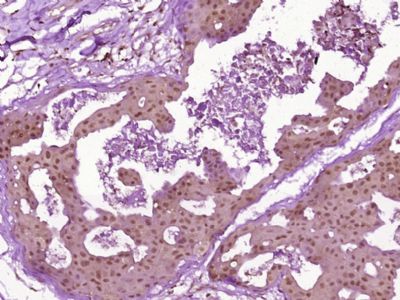
Paraformaldehyde-fixed, paraffin embedded (Human breast carcinoma); Antigen retrieval by boiling in sodium citrate buffer (pH6.0) for 15min; Block endogenous peroxidase by 3% hydrogen peroxide for 20 minutes; Blocking buffer (normal goat serum) at 37°C for 30min; Antibody incubation with (CHK2) Polyclonal Antibody, Unconjugated (SL1391R) at 1:400 overnight at 4°C, followed by operating according to SP Kit(Rabbit) (sp-0023) instructionsand DAB staining.
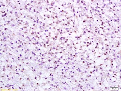
Tissue/cell: human gastric carcinoma; 4% Paraformaldehyde-fixed and paraffin-embedded;
Antigen retrieval: citrate buffer ( 0.01M, pH 6.0 ), Boiling bathing for 15min; Block endogenous peroxidase by 3% Hydrogen peroxide for 30min; Blocking buffer (normal goat serum,SLC0005) at 37℃ for 20 min;
Incubation: Anti-CHK2 Polyclonal Antibody, Unconjugated(SL1391R) 1:200, overnight at 4°C, followed by conjugation to the secondary antibody(SP-0023) and DAB(SLC0010) staining
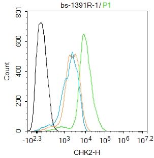
Blank control: A431.
Primary Antibody (green line): Rabbit Anti-CHK2 antibody (SL1391R)
Dilution: 1ug/Test;
Secondary Antibody : Goat anti-rabbit IgG-FITC
Dilution: 0.5ug/Test.
Protocol
The cells were fixed with 4% PFA (10min at room temperature)and then permeabilized with 90% ice-cold methanol for 20 min at -20℃.The cells were then incubated in 5%BSA to block non-specific protein-protein interactions for 30 min at room temperature .Cells stained with Primary Antibody for 30 min at room temperature. The secondary antibody used for 40 min at room temperature. Acquisition of 20,000 events was performed.
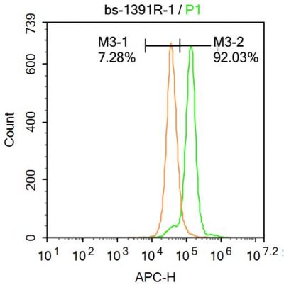
Blank control: A431.
Primary Antibody (green line): Rabbit Anti-CHK2 antibody (SL1391R)
Dilution: 1μg /10^6 cells;
Isotype Control Antibody (orange line): Rabbit IgG .
Secondary Antibody : Goat anti-rabbit IgG-AF647
Dilution: 1μg /test.
Protocol
The cells were fixed with 4% PFA (10min at room temperature)and then permeabilized with 90% ice-cold methanol for 20 min at-20℃. The cells were then incubated in 5%BSA to block non-specific protein-protein interactions for 30 min at at room temperature .Cells stained with Primary Antibody for 30 min at room temperature. The secondary antibody used for 40 min at room temperature. Acquisition of 20,000 events was performed.
Product Feedback Wall
Message :
Your Email :
Copyright © 2007-2018 Sunlong Medical All Rights Reserved.