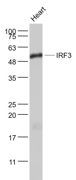
Sample:
Heart (Mouse) Lysate at 40 ug
Primary: Anti- IRF3 (SL2993R) at 1/1000 dilution
Secondary: IRDye800CW Goat Anti-Rabbit IgG at 1/20000 dilution
Predicted band size: 47 kD
Observed band size: 55 kD
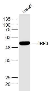
Sample:
Heart (Mouse) Lysate at 40 ug
Primary: Anti-IRF3 (SL2993R) at 1/1000 dilution
Secondary: IRDye800CW Goat Anti-Rabbit IgG at 1/20000 dilution
Predicted band size: 47 kD
Observed band size: 51 kD
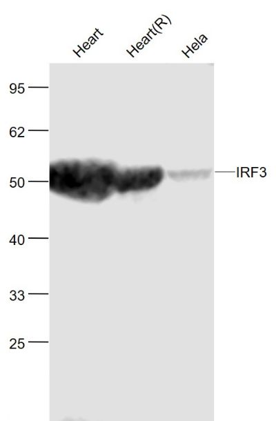
Sample:
Heart(Mouse) Lysate at 40 ug
Heart(Rat) Lysate at 40 ug
Hela(Human) Cell Lysate at 30 ug
Primary: Anti-IRF3 (SL2993R) at 1/1000 dilution
Secondary: IRDye800CW Goat Anti-Rabbit IgG at 1/20000 dilution
Predicted band size: 50 kD
Observed band size: 50 kD
Sample:
Jurkat(Human) Cell Lysate at 30 ug
Panc-1(Human) Cell Lysate at 30 ug
Pancreas (Mouse) Lysate at 40 ug
Kidney (Mouse) Lysate at 40 ug
Spleen (Mouse) Lysate at 40 ug
A431(Human) Cell Lysate at 30 ug
A549(Human) Cell Lysate at 30 ug
Testis (Mouse) Lysate at 40 ug
Lymph node (Mouse) Lysate at 40 ug
Primary: Anti- IRF3 (SL2993R) at 1/1000 dilution
Secondary: IRDye800CW Goat Anti-Rabbit IgG at 1/20000 dilution
Predicted band size: 47 kD
Observed band size: 54 kD
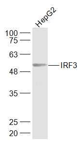
Sample:
Hepg2(Human) Lysate at 40 ug
Primary: Anti-IRF3 (SL2993R) at 1/1000 dilution
Secondary: IRDye800CW Goat Anti-Rabbit IgG at 1/20000 dilution
Predicted band size: 47 kD
Observed band size: 51 kD
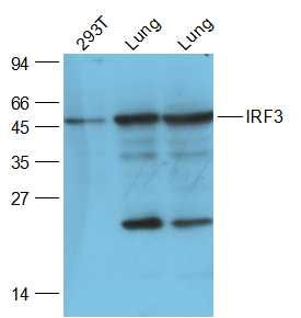
Sample:
293T(Human) Cell Lysate at 30 ug
Lung (Mouse) Lysate at 40 ug
Lung (Rat) Lysate at 40 ug
Primary: Anti-IRF3 (SL2993R) at 1/2000 dilution
Secondary: IRDye800CW Goat Anti-Rabbit IgG at 1/20000 dilution
Predicted band size: 47 kD
Observed band size: 47 kD
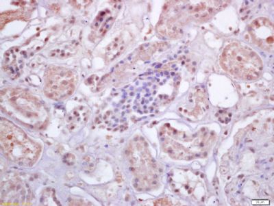
Tissue/cell: human kidney tissue; 4% Paraformaldehyde-fixed and paraffin-embedded;
Antigen retrieval: citrate buffer ( 0.01M, pH 6.0 ), Boiling bathing for 15min; Block endogenous peroxidase by 3% Hydrogen peroxide for 30min; Blocking buffer (normal goat serum,SLC0005) at 37℃ for 20 min;
Incubation: Anti-IRF3 Polyclonal Antibody, Unconjugated(SL2993R) 1:200, overnight at 4°C, followed by conjugation to the secondary antibody(SP-0023) and DAB(SLC0010) staining
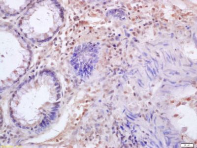
Tissue/cell: human colon carcinoma; 4% Paraformaldehyde-fixed and paraffin-embedded;
Antigen retrieval: citrate buffer ( 0.01M, pH 6.0 ), Boiling bathing for 15min; Block endogenous peroxidase by 3% Hydrogen peroxide for 30min; Blocking buffer (normal goat serum,SLC0005) at 37℃ for 20 min;
Incubation: Anti-IRF3 Polyclonal Antibody, Unconjugated(SL2993R) 1:200, overnight at 4°C, followed by conjugation to the secondary antibody(SP-0023) and DAB(SLC0010) staining
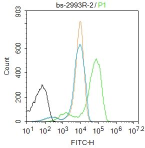
Blank control:Jurkat.
Primary Antibody (green line): Rabbit Anti-IRF3 antibody (SL2993R)
Dilution: 2μg /10^6 cells;
Isotype Control Antibody (orange line): Rabbit IgG .
Secondary Antibody : Goat anti-rabbit IgG-FITC
Dilution: 1μg /test.
Protocol
The cells were fixed with 4% PFA (10min at room temperature)and then permeabilized with 90% ice-cold methanol for 20 min at-20℃. The cells were then incubated in 5%BSA to block non-specific protein-protein interactions for 30 min at room temperature .Cells stained with Primary Antibody for 30 min at room temperature. The secondary antibody used for 40 min at room temperature. Acquisition of 20,000 events was performed.
|