Microtubule-associated proteins (MAPs) regulate microtubule stability and play critical roles in neuronal development and in maintaining the balance between neuronal plasticity and rigidity. MAP-light chain 3 beta (MAP-LC3 Beta) and MAP-light chain 3 alpha (MAP-LC3 Alpha) are subunits of both MAP1A and MAP1B. MAP-LC3 Beta, a homolog of Apg8p, is essential for autophagy and associated to the autophagosome membranes after processing. Two forms of LC3 Beta, the cytosolic LC3-I and the membrane-bound LC3-II, are produced post-translationally. LC3-I is formed by the removal of the SLCterminal 22 amino acids from newly synthesized LC3 Beta, followed by the conversion of a fraction of LC3-I into LC3-II. LC3 enhances fibronectin mRNA translation in ductus arteriosus cells through association with 60S ribosomes and binding to an AU-rich element in the 3’ untranslated region of fibronectin mRNA. This facilitates sorting of fibronectin mRNA onto rough endoplasmic reticulum and translation. MAP LC3 Beta may also be involved in formation of autophagosomal vacuoles. It is expressed primarily in heart, testis, brain and skeletal muscle.
Function:
Probably involved in formation of autophagosomal vacuoles
Subunit:
3 different light chains, LC1, LC2 and LC3, can associate with MAP1A and MAP1B proteins. Interacts at microtubules with CABP1 (via EF-hands 1 and 2) but not with calmodulin. Interacts with FYCO1 (via SLCterminus). Interacts with TP53INP1 and TP53INP2. Interacts with TBC1D25. Directly interacts with SQSTM1; this interaction leads to MAP1LC3B recruitment to inclusion bodies containing polyubiquitinated protein aggregates and to inclusion body degradation by autophagy.
Subcellular Location:
Cytoplasm, cytoskeleton. Endomembrane system; Lipid-anchor. Cytoplasmic vesicle, autophagosome membrane; Lipid-anchor. Note=LC3-II binds to the autophagic membranes.
Tissue Specificity:
Most abundant in heart, brain, skeletal muscle and testis. Little expression observed in liver.
Post-translational modifications:
The precursor molecule is cleaved by APG4B/ATG4B to form LC3-I. This is activated by APG7L/ATG7, transferred to ATG3 and conjugated to phospholipid to form LC3-II.
Similarity:
Belongs to the MAP1 LC3 family.
SWISS:
Q9GZQ8
Gene ID:
81631
Database links:
Entrez Gene: 81631 Human
Entrez Gene: 427559 Chicken
Entrez Gene: 408001 Cow
Entrez Gene: 67443 Mouse
Entrez Gene: 64862 Rat
Omim: 609604 Human
SwissProt: O41515 Cow
SwissProt: Q9GZQ8 Human
SwissProt: Q9CQV6 Mouse
SwissProt: Q62625 Rat
Unigene: 356061 Human
Unigene: 28357 Mouse
Unigene: 41412 Rat
| Picture |
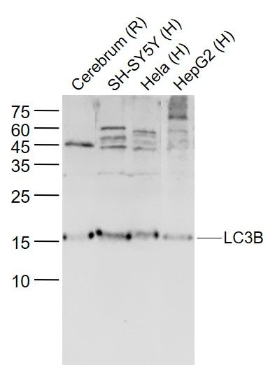
Sample:
Lane 1: Cerebrum (Rat) Lysate at 40 ug
Lane 2: SH-SY5Y (Human) Cell Lysate at 30 ug
Lane 3: Hela (Human) Cell Lysate at 30 ug
Lane 4: HepG2 (Human) Cell Lysate at 30 ug
Primary: Anti-LC3B (SL4843R) at 1/1000 dilution
Secondary: IRDye800CW Goat Anti-Rabbit IgG at 1/20000 dilution
Predicted band size: 14/17 kD
Observed band size: 17 kD
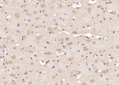
Paraformaldehyde-fixed, paraffin embedded (rat brain); Antigen retrieval by boiling in sodium citrate buffer (pH6.0) for 15min; Block endogenous peroxidase by 3% hydrogen peroxide for 20 minutes; Blocking buffer (normal goat serum) at 37°C for 30min; Antibody incubation with (LC3B) Polyclonal Antibody, Unconjugated (SL4843R) at 1:200 overnight at 4°C, followed by operating according to SP Kit(Rabbit) (sp-0023) instructionsand DAB staining.
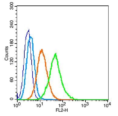
Blank control(blue): TM4 cells(fixed with 2% paraformaldehyde (10 min) , then permeabilized with 90% ice-cold methanol for 30 min on ice).
Primary Antibody:Rabbit Anti-LC3B antibody(SL4843R), Dilution: 1μg in 100 μL 1X PBS containing 0.5% BSA;
Isotype Control Antibody: Rabbit IgG(orange) ,used under the same conditions );
Secondary Antibody: Goat anti-rabbit IgG-PE(white blue), Dilution: 1:200 in 1 X PBS containing 0.5% BSA.
|
|
|