Sample:
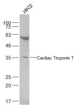
H9C2(Rat) Cell Lysate at 30 ug
Primary: Anti-alpha smooth muscle Actin (SL10648R) at 1/500 dilution
Secondary: IRDye800CW Goat Anti-Rabbit IgG at 1/20000 dilution
Predicted band size: 36 kD
Observed band size: 36 kD
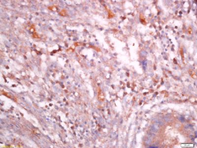
Tissue/cell: human colon cancer; 4% Paraformaldehyde-fixed and paraffin-embedded;
Antigen retrieval: citrate buffer ( 0.01M, pH 6.0 ), Boiling bathing for 15min; Block endogenous peroxidase by 3% Hydrogen peroxide for 30min; Blocking buffer (normal goat serum,SLC0005) at 37℃ for 20 min;
Incubation: Anti-TNNT2 Polyclonal Antibody, Unconjugated(SL10648R) 1:400, overnight at 4°C, followed by conjugation to the secondary antibody(SP-0023) and DAB(SLC0010) staining
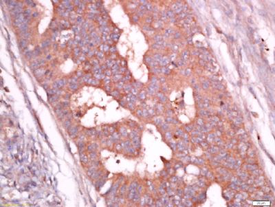
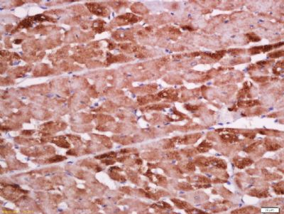
Tissue/cell: rat heart tissue; 4% Paraformaldehyde-fixed and paraffin-embedded;
Antigen retrieval: citrate buffer ( 0.01M, pH 6.0 ), Boiling bathing for 15min; Block endogenous peroxidase by 3% Hydrogen peroxide for 30min; Blocking buffer (normal goat serum,SLC0005) at 37℃ for 20 min;
Incubation: Anti-TNNT2 Polyclonal Antibody, Unconjugated(SL10648R) 1:500, overnight at 4°C, followed by conjugation to the secondary antibody(SP-0023) and DAB(SLC0010) staining
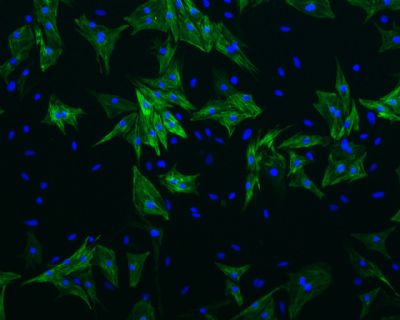
Cell: Neonatal rat ventricular cardiomyocytes;
Dilution: 1:400;
Incubation: Anti-Cardiac Troponin T Antibody, unconjugated (SL10648R);
DAPI was used to stain the cell nuclei.
The image is provided by Tongji University.
Cell: Neonatal rat ventricular cardiomyocytes;
Dilution: 1:400;
Incubation: Anti-Cardiac Troponin T Antibody, unconjugated (SL10648R);
DAPI was used to stain the cell nuclei.
The image is provided by Tongji University.
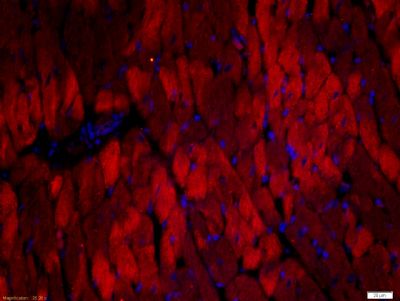
Tissue/cell: rat heart tissue;4% Paraformaldehyde-fixed and paraffin-embedded;
Antigen retrieval: citrate buffer ( 0.01M, pH 6.0 ), Boiling bathing for 15min; Blocking buffer (normal goat serum,SLC0005) at 37℃ for 20 min;
Incubation: Anti-TNNT2 Polyclonal Antibody, Unconjugated(SLR) 1:500, overnight at 4°C; The secondary antibody was Goat Anti-Rabbit IgG, Cy3 conjugated(SL0295G-Cy3)used at 1:200 dilution for 40 minutes at 37°C. DAPI(5ug/ml,blue,SLC0033) was used to stain the cell nuclei
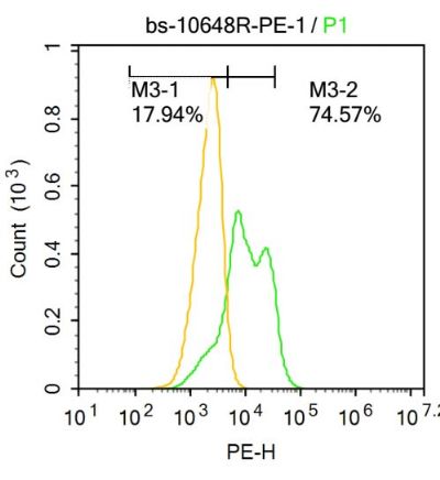
Blank control:U-2OS.
Primary Antibody (green line): Rabbit Anti-TNNT2 antibody (SL10648R)
Dilution: 1μg /10^6 cells;
Isotype Control Antibody (orange line): Rabbit IgG .
Secondary Antibody : Goat anti-rabbit IgG-AF647
Dilution: 1μg /test.
Protocol
The cells were fixed with 4% PFA (10min at room temperature)and then permeabilized with 0.1% PBST for 20 min at-20℃. The cells were then incubated in 5%BSA to block non-specific protein-protein interactions for 30 min at at room temperature .Cells stained with Primary Antibody for 30 min at room temperature. The secondary antibody used for 40 min at room temperature. Acquisition of 20,000 events was performed.
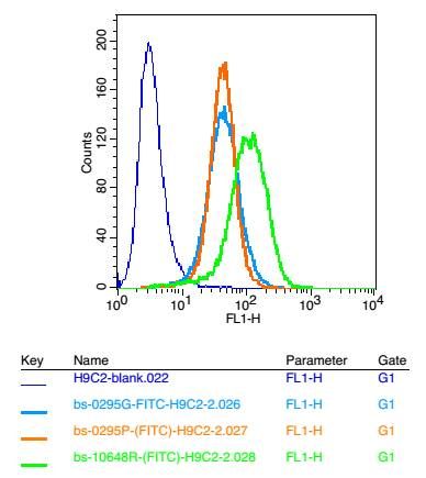
Positive control: H9C2 cells
Concebtration: 2μg/10^6 cells
Incubation conditions: Avoid light , 30 minutes on the ice.
|