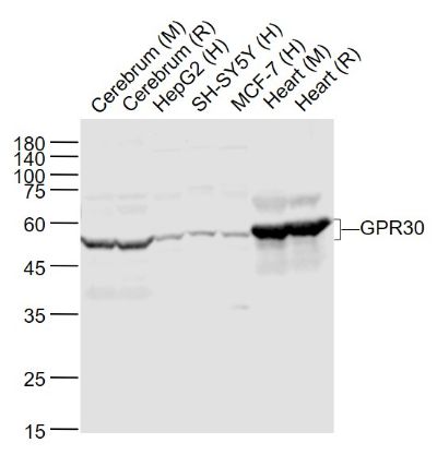
Sample:
Lane 1: Cerebrum (Mouse) Lysate at 40 ug
Lane 2: Cerebrum (Rat) Lysate at 40 ug
Lane 3: HepG2 (Human) Cell Lysate at 30 ug
Lane 4: SH-SY5Y (Human) Cell Lysate at 30 ug
Lane 5: MCF-7 (Human) Cell Lysate at 30 ug
Lane 6: Heart (Mouse) Lysate at 40 ug
Lane 7: Heart (Rat) Lysate at 40 ug
Primary: Anti-GPR30 (SL20643R) at 1/1000 dilution
Secondary: IRDye800CW Goat Anti-Rabbit IgG at 1/20000 dilution
Predicted band size: 55 kD
Observed band size: 55 kD
Sample:
Spleen (Mouse) Lysate at 40 ug
Primary: Anti-GPR30 (SL20643R) at 1/300 dilution
Secondary: IRDye800CW Goat Anti-Rabbit IgG at 1/20000 dilution
Predicted band size: 42 kD
Observed band size: 57 kD
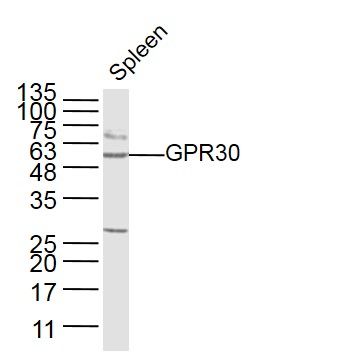
Sample:
Spleen (Mouse) Lysate at 40 ug
Primary: Anti-GPR30 (SL20643R) at 1/300 dilution
Secondary: IRDye800CW Goat Anti-Rabbit IgG at 1/20000 dilution
Predicted band size: 42 kD
Observed band size: 57 kD
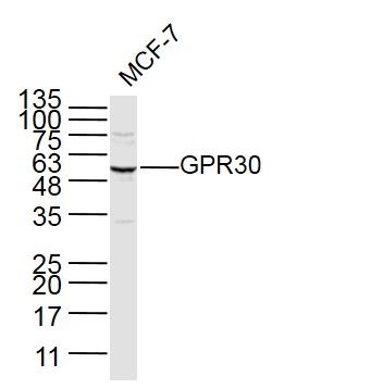
Sample:
MCF-7 (Human) Lysate at 30 ug
Primary: Anti-GPR30 (SL20643R) at 1/300 dilution
Secondary: IRDye800CW Goat Anti-Rabbit IgG at 1/20000 dilution
Predicted band size: 42 kD
Observed band size: 57 kD
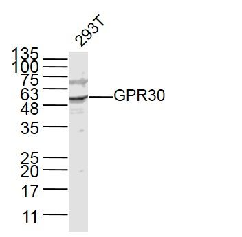
Sample:
293T (Human) Lysate at 30 ug
Primary: Anti-GPR30 (SL20643R) at 1/300 dilution
Secondary: IRDye800CW Goat Anti-Rabbit IgG at 1/20000 dilution
Predicted band size: 42 kD
Observed band size: 57 kD
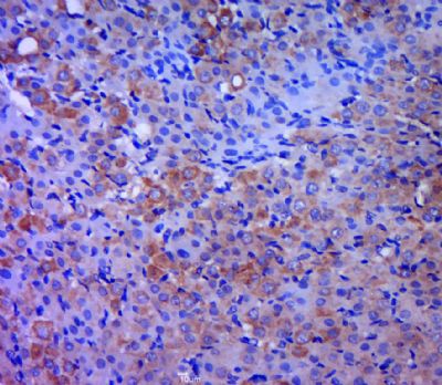
Paraformaldehyde-fixed, paraffin embedded (rat ovary tissue); Antigen retrieval by boiling in sodium citrate buffer (pH6.0) for 15min; Block endogenous peroxidase by 3% hydrogen peroxide for 20 minutes; Blocking buffer (normal goat serum) at 37°C for 30min; Antibody incubation with (GPR30) Polyclonal Antibody, Unconjugated (SL20643R) at 1:400 overnight at 4°C, followed by a conjugated secondary (sp-0023) for 20 minutes and DAB staining.
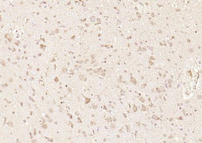
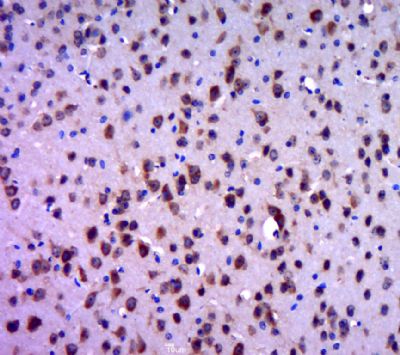
Paraformaldehyde-fixed, paraffin embedded (rat brain); Antigen retrieval by boiling in sodium citrate buffer (pH6.0) for 15min; Block endogenous peroxidase by 3% hydrogen peroxide for 20 minutes; Blocking buffer (normal goat serum) at 37°C for 30min; Antibody incubation with (GPR30) Polyclonal Antibody, Unconjugated (SL20643R) at 1:200 overnight at 4°C, followed by operating according to SP Kit(Rabbit) (sp-0023) instructionsand DAB staining.
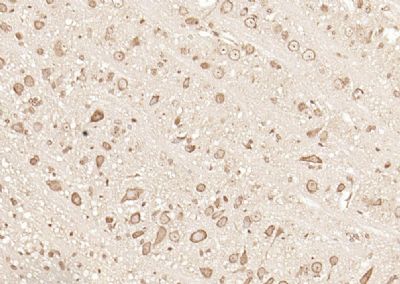
Paraformaldehyde-fixed, paraffin embedded (mouse brain); Antigen retrieval by boiling in sodium citrate buffer (pH6.0) for 15min; Block endogenous peroxidase by 3% hydrogen peroxide for 20 minutes; Blocking buffer (normal goat serum) at 37°C for 30min; Antibody incubation with (GPR30) Polyclonal Antibody, Unconjugated (SL20643R) at 1:200 overnight at 4°C, followed by operating according to SP Kit(Rabbit) (sp-0023) instructionsand DAB staining.
Paraformaldehyde-fixed, paraffin embedded (mouse brain tissue); Antigen retrieval by boiling in sodium citrate buffer (pH6.0) for 15min; Block endogenous peroxidase by 3% hydrogen peroxide for 20 minutes; Blocking buffer (normal goat serum) at 37°C for 30min; Antibody incubation with (GPR30) Polyclonal Antibody, Unconjugated (SL20643R) at 1:400 overnight at 4°C, followed by a conjugated secondary (sp-0023) for 20 minutes and DAB staining.
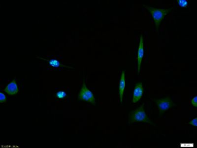
Tissue/cell:SH-SY5Y cell; 4% Paraformaldehyde-fixed; Triton X-100 at room temperature for 20 min; Blocking buffer (normal goat serum,SLC0005) at 37°C for 20 min; Antibody incubation with (GPR30) polyclonal Antibody, Unconjugated (SL20643R) 1:100, 90 minutes at 37°C; followed by a FITC conjugated Goat Anti-Rabbit IgG antibody at 37°C for 90 minutes, DAPI (blue, C02-04002) was used to stain the cell nuclei.
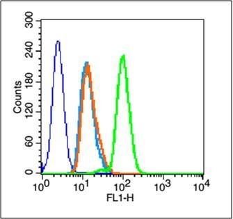
Blank control (blue line): A431 cells (fixed with 70% methanol (Overnight at 4℃) and then permeabilized with 90% ice-cold methanol for 20 min at -20℃).
Primary Antibody (green line): Rabbit Anti-GPR30 antibody (SL20643R),Dilution: 1μg /10^6 cells;
Isotype Control Antibody (orange line): Rabbit IgG .
Secondary Antibody (white blue line): Goat anti-rabbit IgG-FITC,Dilution: 1μg /test.
|