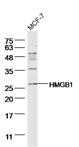
Sample:
MCF-7 Cell (human) Lysate at 40 ug
Primary: Anti- HMGB1 (SL20633R)at 1/300 dilution
Secondary: IRDye800CW Goat Anti-Rabbit IgG at 1/20000 dilution
Predicted band size: 25kD
Observed band size: 27 kD
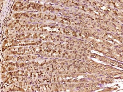
Paraformaldehyde-fixed, paraffin embedded (Rat stomach); Antigen retrieval by boiling in sodium citrate buffer (pH6.0) for 15min; Block endogenous peroxidase by 3% hydrogen peroxide for 20 minutes; Blocking buffer (normal goat serum) at 37°C for 30min; Antibody incubation with (HMGB1) Polyclonal Antibody, Unconjugated (SL20633R) at 1:500 overnight at 4°C, followed by a conjugated secondary (sp-0023) for 20 minutes and DAB staining.
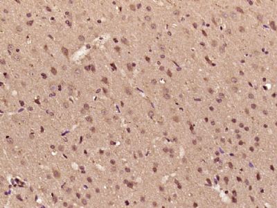
Paraformaldehyde-fixed, paraffin embedded (Rat brain); Antigen retrieval by boiling in sodium citrate buffer (pH6.0) for 15min; Block endogenous peroxidase by 3% hydrogen peroxide for 20 minutes; Blocking buffer (normal goat serum) at 37°C for 30min; Antibody incubation with (HMGB1) Polyclonal Antibody, Unconjugated (SL20633R) at 1:500 overnight at 4°C, followed by a conjugated secondary (sp-0023) for 20 minutes and DAB staining.
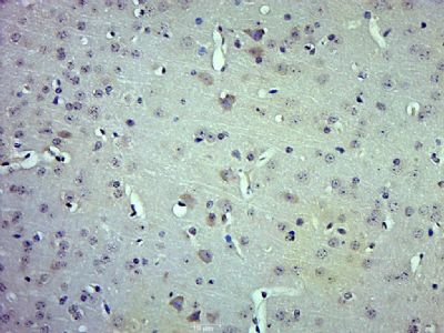
Paraformaldehyde-fixed, paraffin embedded (Mouse brain); Antigen retrieval by boiling in sodium citrate buffer (pH6.0) for 15min; Block endogenous peroxidase by 3% hydrogen peroxide for 20 minutes; Blocking buffer (normal goat serum) at 37°C for 30min; Antibody incubation with (HMGB1) Polyclonal Antibody, Unconjugated (SL20633R) at 1:500 overnight at 4°C, followed by a conjugated secondary (sp-0023) for 20 minutes and DAB staining.
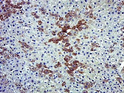
Paraformaldehyde-fixed, paraffin embedded (Rat liver); Antigen retrieval by boiling in sodium citrate buffer (pH6.0) for 15min; Block endogenous peroxidase by 3% hydrogen peroxide for 20 minutes; Blocking buffer (normal goat serum) at 37°C for 30min; Antibody incubation with (HMGB1) Polyclonal Antibody, Unconjugated (SL20633R) at 1:500 overnight at 4°C, followed by a conjugated secondary (sp-0023) for 20 minutes and DAB staining.
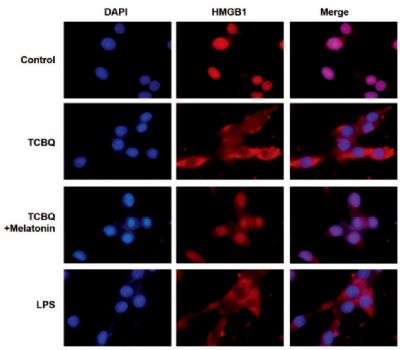
This image was generously provided by Juanli Fu, at Southwest University in Chong Qing, China. 4% Paraformaldehyde fixed PC12 cells stained with Rabbit Anti- HMGB1 Polyclonal Antibody (SL20633R) at 1:300 for 3 hours at 4°C, followed by Rhodamine-conjugated secondary antibody for an additional hour.
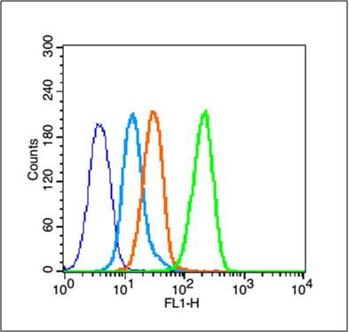
Blank control (blue line): MCF7 (fixed with 80% ethanol (Overnight at 4℃) and then permeabilized with 90% ice-cold methanol for 30 min on ice).
Primary Antibody (green line): Rabbit Anti-HMGB1 antibody (SL20633R),Dilution: 1μg /10^6 cells;
Isotype Control Antibody (orange line): Rabbit IgG .
Secondary Antibody (white blue line): Goat anti-rabbit IgG-FITC,Dilution: 1μg /test.
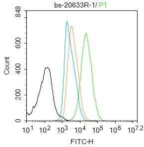
Blank control:HL-60.
Primary Antibody (green line): Rabbit Anti-HMGB1 antibody (SL20633R)
Dilution: 1μg /10^6 cells;
Isotype Control Antibody (orange line): Rabbit IgG .
Secondary Antibody : Goat anti-rabbit IgG-AF488
Dilution: 1μg /test.
Protocol
The cells were fixed with 4% PFA (10min at room temperature)and then permeabilized with 90% ice-cold methanol for 20 min at-20℃. The cells were then incubated in 5%BSA to block non-specific protein-protein interactions for 30 min at room temperature .Cells stained with Primary Antibody for 30 min at room temperature. The secondary antibody used for 40 min at room temperature. Acquisition of 20,000 events was performed.
|