[IF=24.633] Yanxin Li. et al. Decoding the temporal and regional specification of microglia in the developing human brain. Cell Stem Cell. 2022 Mar;: IF ; Mouse,Human.
[IF=2.52] Expression of Pref-1 and Related Chemokines during theDevelopment of Rat Mesenteric Lymph Nodes.(2018)Biomed Environ Sci.ul;31(7):507-514. IHC ; Rat.
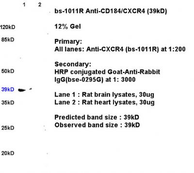
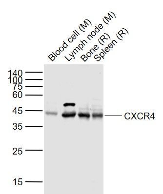
[IF=2.311] Toyoma S et al. SDF-1/CXCR4 induces cell invasion through CD147 in squamous cell carcinoma of the hypopharynx. Oncol Lett. 2020 Aug;20(2):1817-1823. WB ; Rat&Mouse.
[IF=3.457] Hu A et al. Involvement of stromal cell-derived factor-1α (SDF-1α), stem cell factor (SCF), fractalkine (FKN) and VEGF in TSG protection against intimal hyperplasia in rat balloon injury. Biomed Pharmacother. 2019 Feb;110:887-894. IHSLCP ; Rat.
[IF=2.959] Zhuang M et al. Reelin regulates male mouse reproductive capacity via the sertoli cells.(2018) J. Cell. Biochem. 2018 Oct 18 WB ; Mouse.
[IF=13.965] Colas,et al.Impaired Production and Diurnal Regulation of Vascular RvDn-3 DPA Increase Systemic Inflammation and Cardiovascular Disease.(2018) Circulation Research. 122:855-863. FCM ; Human.
[IF=5.878] Jolly,et al.Targeted endothelial gene deletion of Triggering Receptor Expressed on Myeloid cells-1 protects mice during septic shock.(2018) Cardiovascular Research. 114:907-918. IF(IHSLCF) + IF(ICC) ; Mouse + Human.
[IF=3.39] Liu et al. CXCR4-overexpressing bone marrow-derived mesenchymal stem cells improve repair of acute kidney injury. (2013) Am.J.Physiol.Renal.Physio. 305:F1064-73 WB ; Mouse.
[IF=3.3] Ochi, Akinobu, et al. "MIF-2/D-DT Enhances Proximal Tubular Cell Regeneration Through SLPI and ATF4-dependent Mechanisms." American Journal of Physiology-Renal Physiology (2017). WB ; Mouse.
[IF=2.86] Mori, Miki, et al. "Stromal Cell-Derived Factor-1α Plays a Crucial Role Based on Neuroprotective Role in Neonatal Brain Injury in Rats." International Journal of Molecular Sciences 16.8 (2015): 3618-3632. IHSLCF ; Rat.
[IF=3.33] Wu, Qiang, et al. "B-cell lymphoma 6 protein stimulates oncogenicity of human breast cancer cells." BMC Cancer 14.1 (2014): 418. WB ; Human.
[IF=6.321] Sabrina Spiller. et al. Protease-Triggered Release of Stabilized CXCL12 from Coated Scaffolds in an Ex Vivo Wound Model. Pharmaceutics. 2021 Oct;13(10):1597 IF,FC ; Mouse.
[IF=6.639] Qun Lin. et al. Silencing CTNND1 Mediates Triple-Negative Breast Cancer Bone Metastasis via Upregulating CXCR4/CXCL12 Axis and Neutrophils Infiltration in Bone. Cancers. 2021 Jan;13(22):5703 IHC ; Human.
[IF=8.98] Pei, Guangchang, et al. "Renal Interstitial Infiltration and Tertiary Lymphoid Organ Neogenesis in IgA Nephropathy." Clinical Journal of the American Society of Nephrology (2013): CJN-01150113. IHSLCP ; Human.
[IF=3.33] Yin TC et al. Extracorporeal shock wave-assisted adipose-derived fresh stromal vascular fraction restores the blood flow of critical limb ischemia in rat. Vascul Pharmacol. 2019 Feb;113:57-69. IHF ; Rat.
[IF=3.266] Sheu JJ et al. Therapeutic effects of adipose derived fresh stromal vascular fraction-containing stem cellsversus cultured adipose derived mesenchymal stem cells on rescuing heart function in rat after acute myocardial infarction. Am J Transl Res. 2019 Jan 15;11(1):67-86. eCollection 2019. IHF ; Rat.
[IF=3.923] Sha Y et al. MGF E peptide improves anterior cruciate ligament repair by inhibiting hypoxia‐induced cell apoptosis and accelerating angiogenesis.(2018) J Cell Physiol. 2018 Oct 14. IF ; Rabbit.
[IF=6.375] Zhou,et al.CXCR4 antagonist AMD620 enhances the response of MDA-MB-231 triple-negative breast cancer cells to ionizing radiation.(2018) Cancer Letters. 418:196-203. IHSLCP + WB ; Mouse.
[IF=7.84] Zhuo, Wei, et al. ʺThe CXCL12–CXCR4 Chemokine Pathway: A Novel Axis Regulates Lymphangiogenesis.ʺ Clinical Cancer Research 18.19 (2012): 5387-5398. Human, Mouse.
[IF=4.26] Aerbajinai, Wulin, et al. "Glia Maturation Factor-γ Regulates Monocyte Migration through Modulation of β1-Integrin." Journal of Biological Chemistry291.16 (2016): 8549-8564. WB, FCM ; Human.
[IF=1.43] Wang, Xiao-yan, et al. "AMD620 attenuates MMP-3 and MMP-9 expressions and prevents cartilage degradation in a monosodium iodoacetate-induced rat model of temporomandibular osteoarthritis." Journal of Oral and Maxillofacial Surgery (2016). IHSLCP ; Rat.
[IF=3.26] Mu, Hailong, et al. "PLZF‐Induced Upregulation of CXCR4 Promotes Dairy Goat Male Germline Stem Cell Proliferation by Targeting Mir146a." Journal of Cellular Biochemistry (2015). WB ; Goat.
[IF=0.68] Lu Z, Qi L, Bo XJ, Liu GD, Wang JM, Li G. Expression of CD26 and CXCR4 in prostate carcinoma and its relationship with clinical parameters. CD26 and CXCR4 expression shows correlation with prostate cancer. J Res Med Sci 2013;18:647-52 Human.