[IF=2.59] Tian C et al. Short communication: A high-grain diet entails alteration in nutrient chemosensing of the rumen epithelium in goats. Animal Feed Science and Technology. Volume 262, April 2020, 114410. IHSLCP ;
[IF=3.871] Pengli Wang. et al. The AT1 receptor autoantibody causes hypoglycemia in fetal rats via promoting the STT3A-GLUT1-glucose uptake axis in liver. Mol Cell Endocrinol. 2020 Dec;518:111022 WB,IHC ; Rat.
[IF=3.457] Wang N et al. Fibroblast growth factor 21 improves glucose homeostasis partially via down-regulation of Na+-d-glucose cotransporter SGLT1 in the small intestine.(2019) Biomedicine & Pharmacotherapy, 109, 1070–1077. WB ; Mouse.
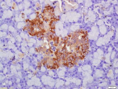
[IF=0] Song B et al. The effects of emodin on insulin resistance in KKAy mice with diabetes mellitus. Phcog Mag. 2018;14:344-50. IHSLCP ; Mouse.
[IF=4.81] Justice,et al.Inhibition of acid sphingomyelinase disrupts LYNUS signaling and triggers autophagy.(2018) Journal of Lipid Research. 59:596-606. IF(ICC) ; Human.
[IF=3.144] Wang,et al.Oat globulin peptides regulate antidiabetic drug targets and glucose transporters in Caco-2 cells.(2018) Journal of Functional Foods. 42:12-20. WB ; Human.
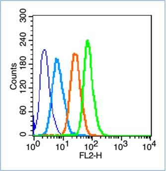
[IF=3.358] Guttman et al. α1-Antitrypsin modifies general NK cell interactions with dendritic cells and specific interactions with islet β-cells in favor of protection from autoimmune diabetes. (2014) Immunology. FC/FACS ; Mouse.
[IF=5.23] Zheng, Liming, et al. "The Modification of Tet1 in Male Germline Stem Cells and Interact with PCNA, HDAC1 to promote their Self-renewal and Proliferation." Scientific Reports 6 (2016): 37414. IF(IHSLCP) ; Goat.
[IF=1.813] Wang Zhe. et al. Total flavonoids of Astragalus Ameliorated Bile Acid Metabolism Dysfunction in Diabetes Mellitus. Evid-Based Compl Alt. 2021;2021:6675567 WB,IF ; Human, Mouse.
[IF=3.571] Jiang T et al. Protein-bound Anthocyanin Compounds of Purple Sweet Potato (p-BASLCPSP) Ameliorate Hyperglycemia by Regulating Hepatic Glucose Metabolism in High Fat Diet/Streptozotocin-Induced Diabetic Mice. J Agric Food Chem. 2020 Feb 12;68(6):1596-1608. WB ; Mouse.