[IF=5.076] Hu C et al. Placentae for Low Birth Weight Piglets Are Vulnerable to Oxidative Stress, Mitochondrial Dysfunction, and Impaired Angiogenesis. Oxid Med Cell Longev. 2020 May 25;2020:8715412. WB ; Pig.
[IF=3.064] Zhao, Ying. et al. Apigenin increases radiosensitivity of glioma stem cells by attenuating HIF-1α-mediated glycolysis. Med Oncol. 2021 Nov;38(11):1-10 WB ; human.
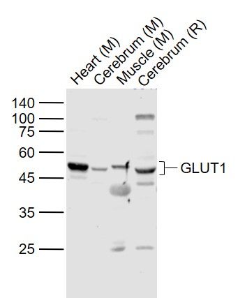
[IF=3.166] Yun S et al. Effects of lead exposure on brain glucose metabolism and insulin signaling pathway in the hippocampus of rats. Toxicol Lett. 2019 Aug;310:23-30. WB ; Rat.
[IF=3.86] Chu, Meiqiang, et al. "MicroRNA-126 participates in lipid metabolism in mammary epithelial cells." Molecular and Cellular Endocrinology (2017). WB ; Human.
[IF=6.543] Chen Mengyuan. et al. Celastrol Protects against Cerebral Ischemia/Reperfusion Injury in Mice by Inhibiting Glycolysis through Targeting HIF-1α/PDK1 Axis. Oxid Med Cell Longev. 2022;2022:7420507 WB ; Mouse.
[IF=5.47] Yan, Yu-E., et al. ʺSignificant Reduction of the GLUT3 Level, but not GLUT1 Level, Was Observed in the Brain Tissues of Several Scrapie Experimental Animals and Scrapie-Infected Cell Lines.ʺ Molecular neurobiology (2013): 1-14. WB ;
[IF=0] Klasvogt, Sonja, et al. "Air–liquid interface enhances oxidative phosphorylation in intestinal epithelial cell line IPESLCJ2." Cell Death Discovery 3 (2017): 17001. WB ; Pig.