[IF=2.15] Zeng, Chao, et al. "Prostate-specific membrane antigen: a new potential prognostic marker of osteosarcoma." Medical Oncology 29.3 (2012): 2234-2239. IHSLCP ; Human.
[IF=9.776] Ruijing Zhao. et al. Inhalation of MSSLCEVs is a noninvasive strategy for ameliorating acute lung injury. J Control Release. 2022 May;345:214 FC ; Human.
[IF=3.352] Qi Wang. et al. Telocytes in The Esophageal Wall of Chickens: A Tale of Subepithelial Telocytes. Poultry Sci. 2022 Mar;:101859 IHC ; Chicken.
[IF=2.744] Cai-Gui Yu. et al. Ultrasound-targeted cationic microbubbles combined with the NFκB binding motif increase SDF-1α gene transfection: A protective role in hearts after myocardial infarction. 2022 Mar 24 IHC ; Rabbit.
[IF=1.702] Xia Dai. et al. Effect of Aerobic and Resistance Training on Endothelial Progenitor Cells in Mice with Type 2 Diabetes. Cell Reprogram. 2020 Aug;22(4):189-197 IF ; Mouse.
[IF=5.135] Ci Li. et al. Sustained release of exosomes loaded into polydopamine-modified chitin conduits promotes peripheral nerve regeneration in rats. Neural Regen Res. 2022 Feb;17(9):2050 FC ; Rat.
[IF=2.175] Zhu, Kun. et al. Effect of lentivirus-mediated growth and differentiation factor-5 transfection on differentiation of rabbit nucleus pulposus mesenchymal stem cells. Eur J Med Res. 2022 Dec;27(1):1-8 FC ; Rabbit.
[IF=7.328] Dongdong Yao. et al. Matrix stiffness regulates bone repair by modulating 12-lipoxygenase-mediated early inflammation. Mat Sci Eng SLCMater. 2021 Sep;128:112359 IF ; Mouse.
[IF=3.263] Mahmoud E. Youssef. et al. α7-nAChRs-mediated therapeutic angiogenesis accounts for the advantageous effect of low nicotine doses against myocardial infarction in rats. Eur J Pharmacol. 2021 Mar;:173996 IHC ; Rat.
[IF=10.652] Zhuo Liang. et al. Exosome derived from mesenchymal stem cells mediates hypoxia-specific BMP2 gene delivery and enhances bone regeneration. Chem Eng J. 2021 Oct;422:130084 IF ; Rabbit.
[IF=2.733] Kai-Li Liu et al. Effects of seawater immersion on open traumatic brain injury in rabbit model. Brain Res. 2020 Sep 15;1743:146903. IHC/IF ; Rabbit.
[IF=4.61] Yue H et al. Gestational exposure to PM2.5 impairs vascularization of the placenta. Sci Total Environ. 2019 May 15;665:153-161. IHSLCP&WB ; Mouse.
[IF=5.579] Chen et al. Lipoxin A4 and its analogue suppress the tumor growth of transplanted H22 in mice: the role of antiangiogenesis. (2010) Mol.Cancer.The. 9:2164-74 IHSLCF ; Mouse.
[IF=2.81] Sun, Wei, et al. "Adipose-Derived Stem Cells Alleviate Radiation-Induced Muscular Fibrosis by Suppressing the Expression of TGF-1." (2015) Stem Cells International FCM ; Rabbit.
[IF=0.78] Zhang, Q. L., et al. "Expression and localization of the vascular endothelial growth factor and changes of microvessel density during hair follicle development of liaoning cashmere goats." Genet Mol Res 12.12 (2013): 6424-6432. IHSLCP ; Goat.
[IF=4.963] Jin J et al. Exosome secreted from adipose-derived stem cells attenuates diabetic nephropathy by promoting autophagy flux and inhibiting apoptosis in podocyte.Stem Cell Res Ther. 2019 Mar 15;10(1):95. ICF ; Human.
[IF=2.445] Mei X et al. Expression of VEGF, CD73 and their relationship with clinical pathology, microvessel density, and prognosis in renal cell carcinoma. Transl Androl Urol. 2020 Jun;9(3):1366-1373. IHSLCP ; Human.
[IF=6.6] Hsueh-Chun Wang. et al. Restoring Osteochondral Defects through the Differentiation Potential of Cartilage Stem/Progenitor Cells Cultivated on Porous Scaffolds. Cells-Basel. 2021 Dec;10(12):3536 FC ; Human.
[IF=2.447] Lei Zhao. et al. Comparative study on the biological characteristics of menstrual blood‑ and endometrium‑derived endometrial cells. Exp Ther Med. 2021 Dec;22(6):1-9 IHC ; Human.
[IF=3.715] Nan Hou. et al. Tissue-engineered esophagus: recellular esophageal extracellular matrix based on perfusion-decellularized technique and mesenchymal stem cells. Biomed Mater. 2021 Aug;: IHC ; Rabbit.
[IF=1.977] Jianqiang Zhao. et al. In vitro facilitating role of polygonatum sibiricum polysaccharide in osteogenic differentiation of bone marrow mesenchymal stem cells from patients with multiple myeloma. 2021 Apr 23 FC ; Human.
[IF=10.652] Pei Liu. et al. Biphasic CK2.1-coated β-glycerophosphate chitosan/LL37-modified layered double hydroxide chitosan composite scaffolds enhance coordinated hyaline cartilage and subchondral bone regeneration. Chem Eng J. 2021 Aug;418:129531 IHC ; Rabbit.
[IF=4.197] Xuewen Qian. et al. Acidosis induces synovial fibroblasts to release vascular endothelial growth factor via acid-sensitive ion channel 1a. Lab Invest. 2020 Aug;101(3):280-291 IHC ; Human.
[IF=1.329] Ziwei Luo. et al. miR‐203a‐3p promotes loureirin A‐induced hair follicle stem cells differentiation by targeting Smad1. Anat Rec. 2021 Mar;304(3):531-540 IF ; Rat.
[IF=3.998] Gae Won Parket al. Topical cell-free conditioned media harvested from adipose tissue-derived stem cells promote recovery from corneal epithelial defects caused by chemical burns. Sci Rep
. 2020 Jul 24;10(1):12448. FCM ; rat.
[IF=3.485] Bai J et al. Irradiation-induced senescence of bone marrow mesenchymal stem cells aggravates osteogenic differentiation dysfunction via paracrine signaling. Am J Physiol Cell Physiol
. 2020 May 1;318(5):C1005-C1017. FCM ; rat.
[IF=1.702] Dai X et al. Effect of Aerobic and Resistance Training on Endothelial Progenitor Cells in Mice with Type 2 Diabetes. Cell Reprogram
. 2020 Aug;22(4):189-197. IF ; mouse.
[IF=5.546] Huang Z et al. The impact of acute stress disorder on gallbladder interstitial cells of Cajal. J Cell Physiol
. 2020 Apr 24. WB,IF ; rabbit.
[IF=4.627] Zhai L et al. Aerobic and resistance training enhances endothelial progenitor cell function via upregulation of caveolin-1 in mice with type 2 diabetes. Stem Cell Res Ther. 2020 Jan 3;11(1):10. ICF ; Mouse.
[IF=4.963] Chen X et al. Transplantation of oral mucosal epithelial cells seeded on decellularized and lyophilized amniotic membrane for the regeneration of injured endometrium.Stem Cell Res Ther. 2019 Mar 21;10(1):107. IHSLCP ; Rat.
[IF=2.976] Guo H et al. Clinical associations between ASCT2 and p-mTOR in the pathogenesis and prognosis of epithelial ovarian cancer. Oncol Rep. 2018 Dec;40(6):3725-3733. IHSLCP ; Human.
[IF=3.923] Sha Y et al. MGF E peptide improves anterior cruciate ligament repair by inhibiting hypoxia‐induced cell apoptosis and accelerating angiogenesis.(2018) J Cell Physiol. 2018 Oct 14. IF ; Rabbit.
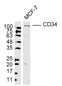
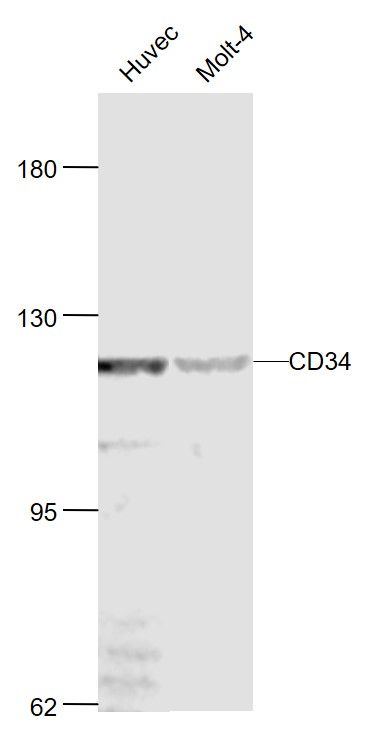
[IF=1.89] Huang et al. Hepatitis B Virus Replication in CD34+ Hematopoietic Stem Cells From Umbilical Cord Blood. (2016) Med.Sci.Monit. 22:1673-81 ICC ; Human.
[IF=2.66] Li et al. Perichondrium mesenchymal stem cells inhibit the growth of breast cancer cells via the DKK-1/Wnt/β-catenin signaling pathway. (2016) Oncol.Rep. 36:936-44 IF(ICC) ; Rat.
[IF=3.427] Gao et al. Common expression of stemness molecular markers and early cardiac transcription factors in human Wharton's jelly-derived mesenchymal stem cells and embryonic stem cells. (2013) Cell.Transplan. 22:1883-900 FCM ; Human.
[IF=2.81] Chang et al. Telocytes in the Spleen. (2015) PLoS.On. 10:e0138851 IF(ICC) ; Rat.
[IF=4.648] Long et al. Mash1-dependent Notch Signaling Pathway Regulates GABAergic Neuron-Like Differentiation from Bone Marrow-Derived Mesenchymal Stem Cells. (2017) Aging.Di. 8:301-313 FCM ; Rat.
[IF=1] Guo, Jian-wen, et al. "Combinatorial effects of Naomai Yihao Capsules () and vascular endothelial growth factor gene-transfected bone marrow mesenchymal stem cells on angiogenesis in cerebral ischemic tissues in rats." Journal of Traditional Chinese Medicine 32.1 (2012): 87-92. IHSLCP ; Rat.
[IF=4.47] Erin, Nuray, et al. "Differential characteristics of heart, liver, and brain metastatic subsets of murine breast carcinoma." Breast Cancer Research and Treatment (2013): 1-13. Mouse.
[IF=0.73] Yu, Z., et al. "Sonic hedgehog and retinoic Acid induce bone marrow-derived stem cells to differentiate into glutamatergic neural cells." Journal of immunoassay & immunochemistry 36.1 (2015): 1. FCM ; Rat.
[IF=1.28] Huang, Sheng‑Li, Jian Jiao, and Hong‑Wei Yan. "Hydrogen-rich saline attenuates steroid-associated femoral head necrosis through inhibition of oxidative stress in a rabbit model." Experimental and therapeutic medicine11.1 (2016): 177-182. IHSLCP ; Rabbit.
[IF=0.94] Bi, Lingyun, et al. "Effect of bone marrow stem cell mobilisation on the expression levels of cellular growth factors in a rat model of acute tubular necrosis." Experimental and Therapeutic Medicine. IHSLCP ; Rat.