DOPA decarboxylase is an enzyme implicated in 2 metabolic pathways, synthesizing 2 important neurotransmitters: dopamine and serotonin which both play key roles in many clinical disorders, including Parkinson's disease. Following the hydroxylation of tyrosine to form L dihydroxyphenylalanine (LDOPA), catalyzed by tyrosine hydroxylase, DDC decarboxylates LDOPA to form dopamine. This neurotransmitter is found in different areas of the brain and is particularly abundant in basal ganglia. Dopamine is also produced by DDC in the sympathetic nervous system and is the precursor of the catecholaminergic hormones, noradrenaline and adrenaline in the adrenal medulla. In the nervous system, tryptophan hydroxylase produces 5 OH tryptophan, which is decarboxylated by DDC, giving rise to serotonin. DDC is a homodimeric, pyridoxal phosphate dependent enzyme.
Function: Catalyzes the decarboxylation of L-3,4-dihydroxyphenylalanine (DOPA) to dopamine, L-5-hydroxytryptophan to serotonin and L-tryptophan to tryptamine.
Subunit: Homodimer.
DISEASE: Defects in DDC are the cause of aromatic L-amino-acid decarboxylase deficiency (AADCD) [MIM:608643]. AADCD deficiency is an inborn error in neurotransmitter metabolism that leads to combined serotonin and catecholamine deficiency. It causes developmental and psychomotor delay, poor feeding, lethargy, ptosis, intermittent hypothermia, gastrointestinal disturbances. The onset is early in infancy and inheritance is autosomal recessive.
Similarity: Belongs to the group II decarboxylase family.
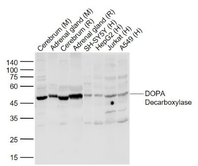
Sample:
Lane 1: Cerebrum (Mouse) Tissue Lysate at 40 ug
Lane 2: adrenal gland (Mouse) TissueLysate at 40 ug
Lane 3: Cerebrum (Rat) Tissue Lysate at 40 ug
Lane 4: adrenal gland (Rat) Tissue Lysate at 40 ug
Lane 5: SH-SY5Y (Human) Cell Lysate at 30 ug
Lane 6: HepG2 (Human) Cell Lysate at 30 ug
Lane 7: Jurkat (Human) Cell Lysate at 30 ug
Lane 8: A549 (Human) Cell Lysate at 30 ug
Primary: Anti-DOPA Decarboxylase (SL036R) at 1/1000 dilution
Secondary: IRDye800CW Goat Anti-Rabbit IgG at 1/20000 dilution
Predicted band size: 53 kD
Observed band size: 50 kD
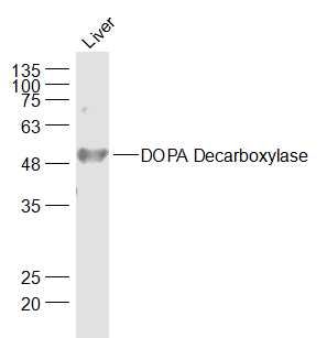
Sample:
Liver (Mouse) Lysate at 40 ug
Primary: Anti-DOPA Decarboxylase (SL036R) at 1/1000 dilution
Secondary: IRDye800CW Goat Anti-Rabbit IgG at 1/20000 dilution
Predicted band size: 53 kD
Observed band size: 53 kD
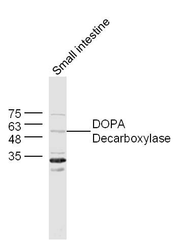
Sample:
Small intestine (Mouse) Lysate at 40 ug
Primary: Anti-DOPA Decarboxylase (SL 036R) at 1/300 dilution
Secondary: IRDye800CW Goat Anti-Rabbit IgG at 1/20000 dilution
Predicted band size: 53 kD
Observed band size: 53 kD
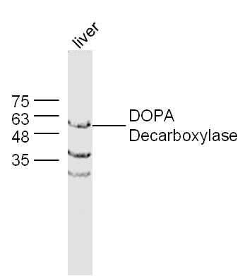
Sample:
Liver (Mouse) Lysate at 40 ug
Primary: Anti-DOPA Decarboxylase (Bs- 036R) at 1/300 dilution
Secondary: IRDye800CW Goat Anti-Rabbit IgG at 1/20000 dilution
Predicted band size: 53 kD
Observed band size: 53 kD
Product Feedback Wall
Specific References (1) | SL036R has been referenced in 1 publications.
[IF=1.29] Hiramoto, Keiichi, Yurika Yamate, and Shosuke Kawanishi. "Detection of Dopa‐positive cells in mouse ovaries in response to ocular exposure to ultraviolet B rays." Photodermatology, Photoimmunology & Photomedicine (2014). IHSLCF ; Mouse.