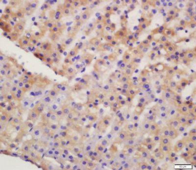
[IF=4.362] WB, IHC ; Mouse.
[IF=1.88] Shihe Zhang. et al. Astragalus polysaccharide regulates brown adipocytes differentiation by miR-6911 targeting Prdm16. 2021 Nov 05 WB ; Mouse.
[IF=5.279] Shuhua Tian. et al. Sulforaphane Regulates Glucose and Lipid Metabolisms in Obese Mice by Restraining JNK and Activating Insulin and FGF21 Signal Pathways. J Agr Food Chem. 2021;XXXX(XXX):XXX-XXX WB ; Mouse.
[IF=2.629] Cao Zhanhong. et al. Protective Effects of Huangqi Shengmai Yin on Type 1 Diabetes-Induced Cardiomyopathy by Improving Myocardial Lipid Metabolism. Evid-Based Compl Alt. 2021;2021:5590623 WB ; Mouse.
[IF=2.146] Xiaolan Wang. et al. The total flavonoids from Selaginella tamariscina (beauv.) Spring improve glucose and lipid metabolism in db/db mice. Iran J Basic Med Sci. 2020 Oct; 23(10): 1286–1292 WB ; Rat.
[IF=4.362] Zhang Yan-hui. et al. α-Lipoic Acid Maintains Brain Glucose Metabolism via BDNF/TrkB/HIF-1α Signaling Pathway in P301S Mice. Front Aging Neurosci. 2020 Aug;12:262 WB ; Mouse.
[IF=2.659] Ye X et al. Irisin reverses insulin resistance in C2C12 cells via the p38-MAPK-PGSLC1α pathway. Peptides. 2019 Jul 24:170120. WB ; Mouse.
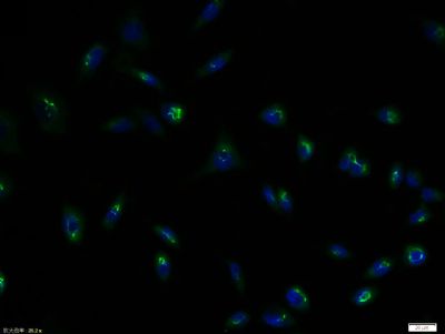
[IF=0] Song B et al. The effects of emodin on insulin resistance in KKAy mice with diabetes mellitus. Phcog Mag. 2018;14:344-50. IHSLCP ; Mouse.
[IF=5.5] Sikder et al. High Fat Diet Upregulates Fatty Acid Oxidation and Ketogenesis via Intervention of PPAR-γ. (2018) Cell.Physiol.Biochem. 48:1317-1331 WB ; Mouse.
[IF=1.58] Gao, Sujie, et al. "Propofol inhibits growth of neurons through regulating insulin receptor and insulin-like growth factor-1 receptor." Int J Clin Exp Pathol 9.7 (2016): 6785-6794. WB ; Rat.
[IF=3.3] Zhang, Shihai, et al. "Effects of isoleucine on glucose uptake through the enhancement of muscular membrane concentrations of GLUT1 and GLUT4 and intestinal membrane concentrations of Na+/glucose co-transporter 1 (SGLT-1) and GLUT2." British Journal of Nutrition (2016): 1-10. WB ; Mouse, Pig.
[IF=5.279] Shuhua Tian. et al. Sulforaphane Regulates Glucose and Lipid Metabolisms in Obese Mice by Restraining JNK and Activating Insulin and FGF21 Signal Pathways. J Agr Food Chem. 2021;XXXX(XXX):XXX-XXX WB ; Mouse.
[IF=3.871] Pengli Wang. et al. The AT1 receptor autoantibody causes hypoglycemia in fetal rats via promoting the STT3A-GLUT1-glucose uptake axis in liver. Mol Cell Endocrinol. 2020 Dec;518:111022 WB,IHC ; Rat.
[IF=3.998] Liza D. Morales. et al. Further evidence supporting a potential role for ADH1B in obesity. Sci Rep-Uk. 2021 Jan;11(1):1-14 WB ; Human.
[IF=3.69] Meng-fan Peng. et al. Effects of total flavonoids from Eucommia ulmoides Oliv. leaves on polycystic ovary syndrome with insulin resistance model rats induced by letrozole combined with a high-fat diet. J Ethnopharmacol. 2021 Jun;273:113947 WB ; Rat.
[IF=1.984] Pan LL et al. Urinary Metabolomics Study of the Intervention Effect of Hypoglycemic Decoction on Type 2 Diabetes Mellitus Rats Model. Evidence-Based Complementary and Alternative Medicine, 2019, 1–17. WB ; Rat.
[IF=2.302] Li Q et al. All-trans retinoic acid regulates sheep primary myoblasts proliferation and differentiation in vitro. Domestic Animal Endocrinology,2019 106394. WB ; Sheep.
[IF=1.832] Zhang T et al. Dietary Sea Buckthorn Pomace Induces Beige Adipocyte Formation in Inguinal White Adipose Tissue in Lambs. Animals (Basel). 2019 Apr 24;9(4). pii: E193. WB ; ram lambs.